The human heart is an extraordinary organ whose primary function is to pump blood through the body. Beyond its mechanical function of pumping blood, the heart possesses a sophisticated electrical system that coordinates its rhythmic contractions.
Studying how the heart generates and regulates signals can provide valuable insights into heart and overall health. Understanding and interpreting EKG rhythms can offer important information about the body's well being. This knowledge can help in assessing the health of the heart and identifying potential issues early on. Monitoring and interpreting the electrical signals of the heart can be a key factor in maintaining good heart health.
How the Heart Creates Electrical Activity
The heart's electrical activity originates from a group of specialized cells known as the sinoatrial (SA) node, located in the right atrium. Often referred to as the natural pacemaker, the SA node generates electrical impulses at regular intervals. These impulses initiate the contraction of the heart muscles, pumping blood throughout the body.
The electrical impulse from the SA node spreads across the atria. This impulse then causes them to contract and push blood into the ventricles. The impulse then reaches the atrioventricular (AV) node. This acts as a gateway that delays the signal before it passes into the ventricles.
This delay ensures the atria have enough time to empty their blood into the ventricles before the ventricles contract. The impulse passes through the AV node. It then travels along the bundle of His, which splits into the right and left bundle branches.
Finally, the impulse spreads throughout the ventricles via the Purkinje fibers. This orchestrated conduction system ensures that the heartbeats in a coordinated and efficient manner.
Recording the Electrical Activity of the Heart: ECG vs. EKG
Doctors commonly measure the electrical activity of the heart using an electrocardiogram, abbreviated as ECG or EKG (from the German "Elektrokardiogramm"). Both terms are interchangeable and refer to the same diagnostic tool; EKG vs ECG, it doesn't matter.
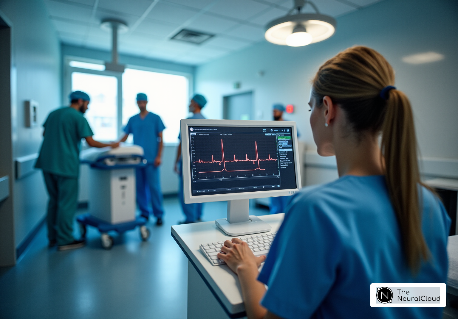
An ECG/EKG involves placing electrodes on the skin at specific points on the body, including the chest, arms, and legs. These electrodes detect the tiny electrical changes on the skin that arise from the heart's electrical activity. The signals amplify, record, and display as ECG waves and ECG strips on a monitor or printed on paper.
An ECG machine can store the data it collects for future reference, analysis, and comparison. Modern devices often come with software that can analyze the EKG strips and store the data. They can also detect abnormal ECGs and integrate with electronic health records for comprehensive patient management.
What the ECG Actually Tells You
An ECG provides a visual representation of the heart's electrical activity over a period of time. The primary components of ECG waves include the P wave, QRS complex, and T wave:
- P Wave: Represents atrial depolarization, which is the electrical activity that triggers the atria to contract.
- QRS Complex: Represents ventricular depolarization, the electrical activity that causes the ventricles to contract. This is the most prominent part of the ECG.
- T Wave: Represents ventricular repolarization. This is the process of the ventricles resetting electrically to prepare for the next contraction.
Healthcare professionals can learn from examining the cardiac rhythm strips, heart rate, and electrical conduction pathways. ECG abnormalities can indicate various cardiac conditions.
ECG Rhythms
We broadly categorize ECG rhythms into normal and abnormal rhythms. The normal rhythm, known as sinus rhythm, originates from the SA node and maintains a regular rate and rhythm. Conditions that deviate from sinus rhythm, result in abnormal ECG; examples include:
- Bradycardia: A slower than normal heart rate, typically less than 60 beats per minute.
- Tachycardia: A faster than normal heart rate, typically more than 100 beats per minute.
- Atrial Fibrillation: An irregular and often rapid heart rate originating from abnormal electrical signals in the atria. This would result in the ECG having arrhythmia.
- Ventricular Fibrillation: A life-threatening condition characterized by chaotic electrical activity in the ventricles, leading to ineffective heart contractions.
- Premature Contractions: Early heartbeats originating from the atria (PACs) or ventricles (PVCs) that disrupt the regular heart rhythm.
What Abnormalities in Electrical Activity Mean for Heart and Overall Health
Abnormalities in the heart's electrical activity, as detected by an ECG, can be indicative of various underlying health issues. Some common abnormalities include:
- Ischemia and Infarction: Reduced blood flow to the heart muscle (ischemia) or a complete blockage (infarction). Changes in the ST segment and T wave can occur, suggesting a heart attack or other coronary artery disease.
- Electrolyte Imbalances: Abnormal levels of potassium, calcium, and magnesium can affect the heart's electrical activity, leading to characteristic changes in the ECG.
- Conduction Block: Delays or blocks in the electrical conduction pathways. Things such as bundle branch blocks or an AV block can slow down or disrupt the normal heartbeat.
- Hypertrophy: Enlarged heart muscles can change ECG rhythms. Conditions like hypertension or cardiomyopathy can cause this and indicate increased cardiac workload. Resistance training can also achieve hypertrophy and beneficially grow the heart muscles through exercise.
How AI and Neural Networks can benefit ECG Analysis
Neural Cloud Solutions Inc. has developed a Neural Network for labeling ECGs that could revolutionize how heart conditions are diagnosed and interpreted. It can provide rapid, accurate, beat-by-beat analysis of any length ECG within minutes. This advanced AI technology can process and label each heartbeat's data points within minutes. This significantly reduces the time required for manual interpretation by healthcare professionals.
The ECG labeling Neural Network, MaxYield™, can help detect normal and abnormal heart rhythms. It can also detect subtle changes in the P wave, QRS complex, and T wave. Additionally, it can support recognition of patterns indicative of heart conditions like atrial fibrillation, ventricular tachycardia, or myocardial ischemia.
Our MaxYield Neural Network helps enhance diagnostic accuracy and efficiency. This allows for quicker medical intervention, personalized treatment plans, and improved patient outcomes, especially in critical scenarios where timely diagnosis is crucial. The consistent and objective analysis provided by MaxYield minimizes human error. It supports continuous monitoring and real-time risk stratification in various healthcare settings.
Conclusion
The heart's electrical activity is a critical aspect of its function, allowing for coordinated contractions that sustain life. Healthcare providers use ECG for measuring and analyzing heart activity. This helps them understand the heart's rhythm and identify any ECG abnormalities that may indicate serious health issues.
Understanding EKG rhythm strips and what they reveal about the heart's health can aid in early diagnosis. This leads to effective treatment and improved outcomes for patients with cardiovascular issues.
With Neural Cloud Solutions’ MaxYield platform, the integration of artificial intelligence and machine learning in ECG analysis promises to further enhance our ability to monitor and interpret heart health. This will pave the way for more personalized and precise medical care.


