Introduction
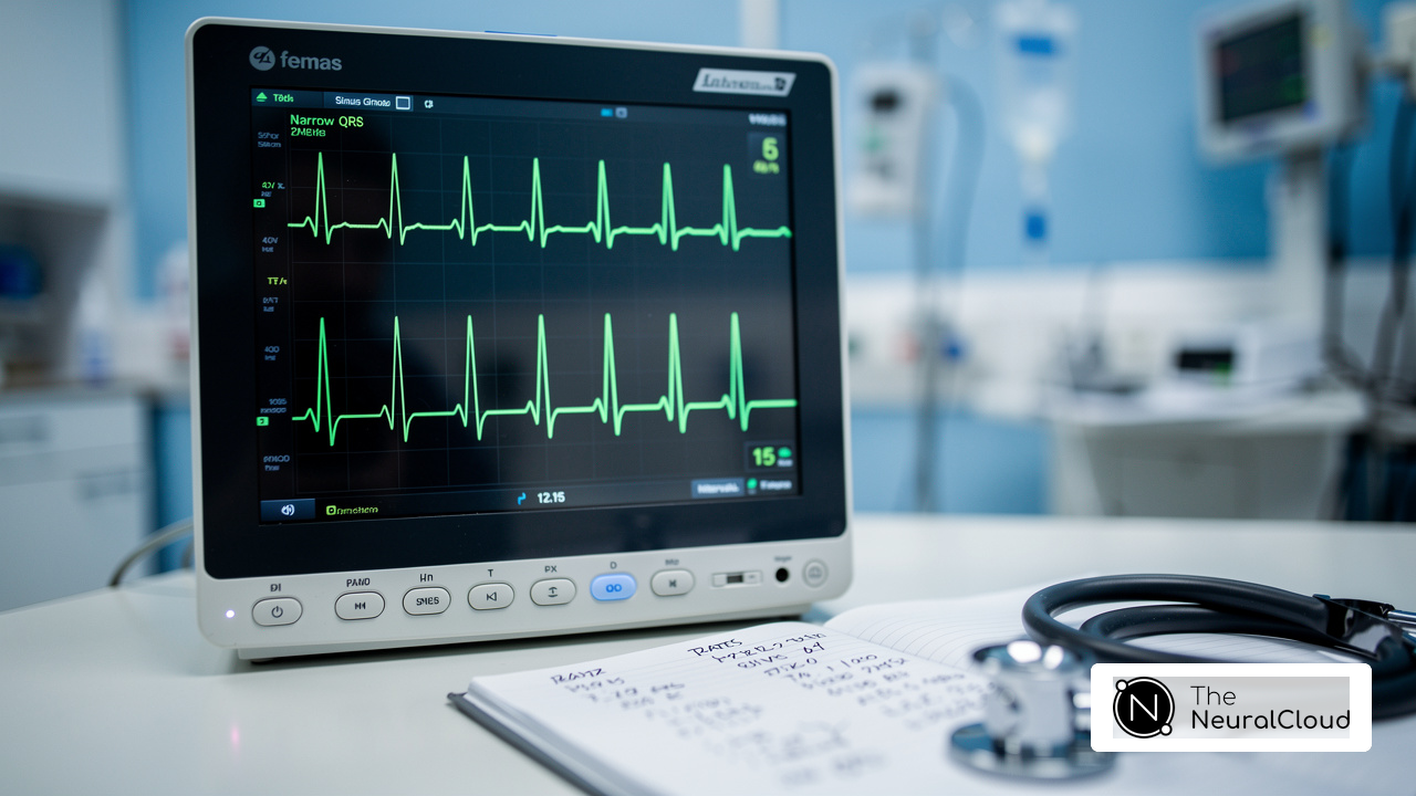
Peaked T waves on an electrocardiogram (ECG) are crucial indicators of potential cardiac issues, including hyperkalemia and myocardial ischemia. However, the complexity of T wave morphology can pose significant challenges for healthcare professionals, complicating clinical interpretations and increasing the risk of misdiagnosis. This is where innovative solutions like MaxYield™ come into play.
MaxYield™ enhances the analysis of T waves through advanced AI technology, streamlining the diagnostic process. By providing detailed insights into T wave morphology, the platform allows clinicians to make quicker and more accurate diagnoses. This capability is particularly beneficial in high-stakes environments where timely decision-making is essential for patient care.
The advantages of utilizing MaxYield™ are clear. Healthcare professionals can leverage its features to navigate the complexities of ECG interpretation more effectively. This not only improves diagnostic accuracy but also enhances patient outcomes, ultimately leading to better healthcare delivery. With tools like MaxYield™, clinicians are better equipped to address the challenges of ECG analysis, ensuring that they can provide the highest standard of care.
Define T Waves: Characteristics and Importance in ECG
of ventricular repolarization during the cardiac cycle, appearing as a positive shift after the QRS complex on an ECG. However, their morphology can vary significantly due to factors like lead placement, heart rate, and underlying cardiac conditions. Typically, T waves have an asymmetrical shape, characterized by a gradual ascent followed by a quicker descent. Statistically, the normal T wave morphology is well-defined, with amplitudes generally less than 5 mm.
Clinicians need to be adept at recognizing variations in T waves, including peaked T waves, as these can signal various cardiac conditions, such as ischemia and electrolyte imbalances. Recent studies highlight the importance of T wave signals in ECG interpretation, revealing that misinterpretations can lead to misdiagnosis. For example, peaked T waves may indicate hyperkalemia, while inverted T patterns can suggest cardiac ischemia.
The technology addresses the challenges posed by physiological variability and signal artifacts in ECG readings. It features advanced noise filtering and distinct signal recognition, enhancing the accuracy of T wave analysis. This ensures that clinicians can make informed decisions based on clearer, more reliable data.
By specifically targeting the nuances of T waves and filtering out irrelevant noise, MaxYield™ significantly improves the interpretative process. This ultimately leads to better patient management, allowing healthcare professionals to provide more accurate diagnoses and treatment plans.

Explore Peaked T Waves: Clinical Implications and Diagnostic Relevance
are characterized by their tall, narrow, and symmetric shape, typically exceeding 5 mm in limb electrodes and 10 mm in precordial electrodes. These fluctuations are primarily associated with hyperkalemia, a condition marked by elevated potassium levels that can lead to serious complications. Recent studies indicate that peaked T waves not only signify hyperkalemia but may also indicate early signs of other cardiac abnormalities. For instance, hyperacute T waves, which are noted for their increased height and symmetry, can be mistaken for T-wave alterations due to hyperkalemia, presenting diagnostic challenges for clinicians. Recognizing these patterns is crucial for timely intervention, as they may precede more severe conditions like acute myocardial infarction.
The diagnostic significance of peaked T waves in hyperkalemia is highlighted by their frequent occurrence in patients with chronic kidney disease, where 67% of cases are attributed to this condition. To address the challenges in diagnosis, integrating AI technology with ECG analysis can significantly enhance efficiency. MaxYield™ automates the labeling and data extraction processes, which reduces operational costs and tackles issues such as physiological variability and signal artifacts. This advanced AI technology transforms lengthy and noisy ECG recordings into clean, crisp signals, allowing clinicians to make informed decisions regarding patient management.
The benefits of using MaxYield™ are substantial for healthcare professionals. By streamlining ECG analysis, the platform not only improves accuracy but also enhances patient outcomes in emergency settings. Key features of MaxYield™ include:
- Automation of labeling and data extraction
- Reduction of operational costs
- Enhanced clarity of ECG signals
In summary, MaxYield™ empowers healthcare professionals to navigate the complexities of ECG interpretation with greater ease, ultimately leading to better patient care.

Analyze Peaked T Waves: Step-by-Step ECG Interpretation Techniques
To effectively analyze T waves, follow these systematic steps:
- Identify the T Component: Locate the T component that follows each QRS complex on the ECG. Ensure that the signal is distinctly visible, free from noise or artifacts that could obscure its features.
- Measure Amplitude: Utilize calipers or digital measurement tools to measure amplitude. Typically, T waves on ECG exceed 5 mm in limb leads and 10 mm in precordial leads, indicating potential underlying issues.
- Assess Morphology: Peaked T waves on ECG should be tall and narrow, characterized by a symmetric peak that differentiates them from other T wave shapes.
- Examine Electrode Positioning: Assess the T deflection across various electrodes to verify if the peaked shape is uniform throughout the ECG. Variability in lead placement can affect interpretation.
- Review Clinical History: Review the patient's clinical history and relevant laboratory results, particularly potassium levels, to assess the importance of the peaked T pattern. This comprehensive approach ensures a thorough interpretation of T abnormalities, facilitating accurate diagnosis.
- Leverage AI Technology: The platform from Neural Cloud Solutions enhances ECG analysis through its advanced algorithms. This platform analyzes T waves on ECG and other abnormalities, providing real-time alerts that significantly improve the urgency of clinical responses. By facilitating timely interventions, it plays a crucial role in emergency settings.
By mastering these techniques, healthcare professionals can enhance their diagnostic capabilities and improve patient outcomes in cardiac care.

Integrate Advanced Technology: Enhancing ECG Analysis with AI Solutions
faces significant challenges, particularly in the precision and efficiency of interpreting T signals. Traditional methods can struggle to identify subtle patterns, which is where AI solutions come into play. The technology leverages advanced algorithms to rapidly analyze extensive ECG data, pinpointing patterns that might escape human detection.
One of the standout features of MaxYield™ is its use of machine learning models, like those found in Neural Cloud Solutions' offerings. These models excel at recognizing abnormalities on ECG and linking them to critical clinical outcomes, such as patient risk stratification. By automating the detection of these abnormalities, healthcare professionals can shift their focus from time-consuming manual analysis to high-level decision-making.
The benefits of using MaxYield™ are clear:
- It streamlines workflows, enabling real-time analysis and facilitating timely interventions in clinical settings.
- Notably, MaxYield™ boasts a diagnostic accuracy significantly higher than the 86.7% sensitivity of traditional physician reporting. This improvement underscores the technology's potential to elevate diagnostic accuracy.
- Moreover, AI models have shown a marked increase in predictive performance, which enhances the reliability of ECG assessments, especially in emergency situations.
With MaxYield™, healthcare professionals can trust that they are equipped with the tools to make informed decisions swiftly.

Conclusion
Mastering the nuances of peaked T waves on ECG is essential for accurate cardiac assessment and timely interventions. This article has explored the significance of T wave morphology, particularly the implications of peaked T waves, which can indicate serious conditions such as hyperkalemia and myocardial ischemia. Understanding these variations is crucial for healthcare professionals to enhance diagnostic accuracy and improve patient outcomes.
In the realm of ECG analysis, challenges often arise due to the complexity of T wave patterns. The MaxYield™ platform addresses these challenges by offering advanced features that streamline ECG interpretation. Its ability to analyze T wave morphology in real-time allows clinicians to quickly identify abnormalities, ensuring timely interventions. This not only enhances diagnostic precision but also supports better patient management in critical situations.
Key features of the MaxYield™ platform include:
- Its user-friendly interface
- Automated analysis capabilities
- Integration of AI technology
These features empower healthcare professionals to interpret ECGs with greater accuracy and efficiency. By leveraging AI solutions, clinicians can detect abnormalities that may otherwise go unnoticed, ultimately leading to improved patient outcomes.
The advantages of utilizing the MaxYield™ platform are significant. Clinicians benefit from reduced analysis time, allowing for quicker decision-making in both emergency and routine settings. Additionally, the platform's advanced algorithms enhance the reliability of ECG interpretations, fostering confidence in clinical decisions. As the landscape of ECG technology evolves, embracing innovations like MaxYield™ will transform patient care, enabling healthcare professionals to make informed, swift decisions.
Frequently Asked Questions
What are T waves in an ECG?
T waves are crucial indicators of ventricular repolarization during the cardiac cycle, appearing as a positive shift after the QRS complex on an ECG.
What is the typical morphology of normal T waves?
Normal T waves typically have an asymmetrical shape, characterized by a gradual ascent followed by a quicker descent.
What are the normal duration and amplitude ranges for T waves?
The normal T duration ranges from 0.10 to 0.25 seconds, with amplitudes generally less than 5 mm.
Why is it important for clinicians to recognize variations in T wave morphology?
Recognizing variations in T wave morphology is important because they can signal various cardiac conditions, such as ischemia and electrolyte imbalances.
What do peaked T waves on an ECG indicate?
Peaked T waves may indicate hyperkalemia or myocardial ischemia.
What do inverted T patterns suggest in an ECG?
Inverted T patterns can suggest cardiac ischemia.
How does the MaxYield™ platform enhance ECG analysis?
The MaxYield™ platform addresses challenges in ECG analysis by featuring advanced noise filtering and distinct signal recognition, which enhances the accuracy of T wave analysis.
What is the benefit of improved T wave analysis using MaxYield™?
Improved T wave analysis using MaxYield™ leads to better patient management by allowing healthcare professionals to make more accurate diagnoses and treatment plans.
List of Sources
- Define T Waves: Characteristics and Importance in ECG
- sciencedirect.com (https://sciencedirect.com/topics/medicine-and-dentistry/t-wave-amplitude)
- ECG T Wave Inversion Features Predict Cardiomyopathy | www.PhysiciansWeekly.com (https://physiciansweekly.com/post/ecg-t-wave-inversion-features-predict-cardiomyopathy)
- AI-ECG Finds STEMI Faster, Cuts False-Positive Cath Lab Activations (https://tctmd.com/news/ai-ecg-finds-stemi-faster-cuts-false-positive-cath-lab-activations)
- skillstat.com (https://skillstat.com/glossary/t-wave)
- Explore Peaked T Waves: Clinical Implications and Diagnostic Relevance
- powerfulmedical.com (https://powerfulmedical.com/blog/hyperacute-t-waves)
- onlinelibrary.wiley.com (https://onlinelibrary.wiley.com/doi/10.1111/nep.70100)
- Unmasking Hyperkalemia: Highlighting Critical ECG Changes (https://powerfulmedical.com/blog/hyperkalemia-ecg-critical-changes)
- link.springer.com (https://link.springer.com/article/10.1186/s12873-019-0247-0)
- sciencedirect.com (https://sciencedirect.com/science/article/pii/S0736467918309235)
- Analyze Peaked T Waves: Step-by-Step ECG Interpretation Techniques
- powerfulmedical.com (https://powerfulmedical.com/blog/hyperacute-t-waves)
- ecg-od.com (https://ecg-od.com/case_studies/royal-brompton-hospitals-use-of-ecg-on-demands-ai-assisted-mail-order-holter-monitoring-service)
- Unmasking Hyperkalemia: Highlighting Critical ECG Changes (https://powerfulmedical.com/blog/hyperkalemia-ecg-critical-changes)
- openheart.bmj.com (https://openheart.bmj.com/content/4/1/e000561)
- theneuralcloud.com (https://theneuralcloud.com/post/master-peaked-t-waves-essential-ecg-analysis-techniques)
- Integrate Advanced Technology: Enhancing ECG Analysis with AI Solutions
- PMcardio Reports Positive RCT Results and Late-Breaking Clinical Science for STEMI Detection (https://powerfulmedical.com/blog/pmcardio-reports-positive-rct-results-and-late-breaking-clinical-science-for-stemi-detection)
- AI-Based ECG Analysis Significantly Improves STEMI Detection, Reduces False Activations - American College of Cardiology (https://acc.org/latest-in-cardiology/articles/2025/10/24/16/56/tues-554pm-ai-tct-2025)
- cureus.com (https://cureus.com/articles/405923-the-use-of-artificial-intelligence-in-ecg-interpretation-in-the-outpatient-setting-a-scoping-review)
- AI dramatically improves the detection of severe heart attacks (https://cardiovascularbusiness.com/topics/artificial-intelligence/ai-dramatically-improves-detection-severe-heart-attacks)
- Artificial Intelligence–Assisted ECG Interpretation versus Conventional Reporting in Predicting Arrhythmias in Acute Coronary Syndrome: A Diagnostic Accuracy Study (https://healthcare-bulletin.co.uk/article/artificial-intelligence-assisted-ecg-interpretation-versus-conventional-reporting-in-predicting-arrhythmias-in-acute-coronary-syndrome-a-diagnostic-accuracy-study-4252)