Introduction
Despite its importance, accurately interpreting ECG changes in myocardial infarction remains a significant challenge for healthcare professionals. Understanding the nuances of myocardial infarction (MI) is crucial for effective cardiac care, particularly as the condition remains a leading cause of mortality in Canada. The electrocardiogram (ECG) serves as a vital tool in diagnosing MI, revealing key changes that can indicate the severity and presence of heart damage.
Clinicians often struggle to decipher the intricate details of ECG readings, which can lead to misdiagnosis or delayed treatment. So, what should clinicians be on the lookout for in ECG readings to act quickly when a patient is having an MI? Understanding these alterations is essential for ensuring timely interventions that can save lives.
Define Myocardial Infarction and Its ECG Significance
Diagnosing myocardial infarction (MI) can be complex, especially when relying solely on traditional methods. Myocardial damage occurs when blood flow to a section of the heart is blocked, leading to harm or death of heart muscle tissue. Blockages in one or more coronary arteries, often due to plaque buildup, frequently cause this condition. Understanding what does an MI look like on an ECG is important because it reveals vital changes that indicate both the presence and severity of the damage. Key ECG changes that illustrate what does an MI look like on an ECG include:
- ST-segment elevation
- T-wave inversions
- Development of pathological Q waves
These alterations are crucial for timely diagnosis and treatment, guiding healthcare professionals in making informed clinical decisions.
In Canada, the prevalence of acute myocardial infarction remains a pressing public health concern, with 51,365 deaths attributed to this condition in the fiscal year 2022-2023. Studies show that AI methods can really boost how we manage suspected acute coronary syndrome, making diagnoses more accurate. For instance, a deep learning model demonstrated superior performance in identifying obstructive MI, achieving an accuracy rate of 84% compared to traditional methods. This shows just how important it is to bring innovative tech into healthcare to improve outcomes for those with MI.
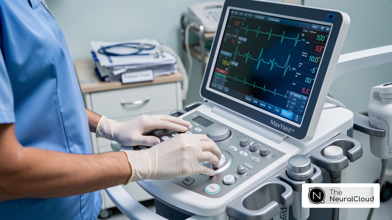
With Neural Cloud Solutions' MaxYield™, these ECG changes can be analyzed with advanced noise filtering and automation, transforming noisy recordings into detailed insights. MaxYield™ delivers beat-by-beat analysis, outputting an analysis of 200,000 heartbeats in less than 5 minutes, enabling healthcare professionals to identify cardiac events swiftly and confidently. Additionally, MaxYield™ is pending FDA Class II SaMD clearance and is designed to integrate seamlessly into clinical workflows. The inclusion of Insight360, a customizable visualization and reporting tool, further enhances the integration of MaxYield™ by transforming clean ECG data into interactive dashboards and clinical-ready reports. This combination not only enhances diagnostic yield but also supports informed clinical decision-making, ultimately improving outcomes for individuals.
Case studies have further illustrated what does an MI look like on an ECG, demonstrating the effectiveness of ECG in diagnosing myocardial infarction. One study examined records from over 1,000 individuals suspected of having a STEMI heart attack, revealing that AI models could accurately identify 553 confirmed cases on the initial ECG, surpassing the 427 detected by traditional methods. These findings highlight the critical role of ECG in early detection and intervention, ultimately saving lives and improving patient care. Incorporating advanced technologies like MaxYield™ could redefine how healthcare professionals approach MI diagnosis and treatment.

Identify Key ECG Features of Myocardial Infarction
Accurate diagnosis of myocardial damage is crucial for timely intervention, yet it remains a challenge due to the subtlety of ECG changes. Understanding what does an MI look like on an ECG is essential for precise diagnosis and prompt intervention regarding myocardial damage. These features include:
- ST-Segment Elevation: This is a primary indicator of an acute MI, especially in ST-Elevation Myocardial Infarction (STEMI). The ST segment is elevated above the baseline in two or more contiguous leads, signaling transmural ischemia. More than 3 million individuals develop STE-MI each year, underscoring the importance of recognizing this feature promptly.
- T-Wave Changes: Initially, T waves may appear hyperacute (tall and peaked) and subsequently invert as the tissue damage progresses. These changes help us understand how the myocardial injury is progressing.
- Pathological Q Waves: These may develop hours to days after the onset of MI, indicating necrosis of myocardial tissue. The presence of Q waves in specific leads can help identify the area of damage, which is crucial for targeted treatment.
- Mutual Adjustments: These changes may occur in leads opposite to the region of tissue death, providing further diagnostic information. Understanding these reciprocal changes can aid in confirming the diagnosis of what does an MI look like on an ECG.
By 2026, advancements in ECG interpretation have significantly improved the identification of these key features, allowing for more precise and timely management of heart attack cases. For instance, studies have shown that AI-based ECG interpretation can significantly improve the identification of obstructive MI, achieving a sensitivity of 77% and specificity of 99%. Furthermore, platforms like MaxYield™ and its visualization tool Insight360 transform clean ECG data into interactive dashboards and clinical-ready reports, highlighting the evolving landscape of cardiac diagnostics and the critical role of ECG in emergency settings. As technology evolves, the ability to interpret ECGs accurately will be paramount in enhancing patient care during critical moments.

Explore the Evolution of MI Diagnosis via ECG
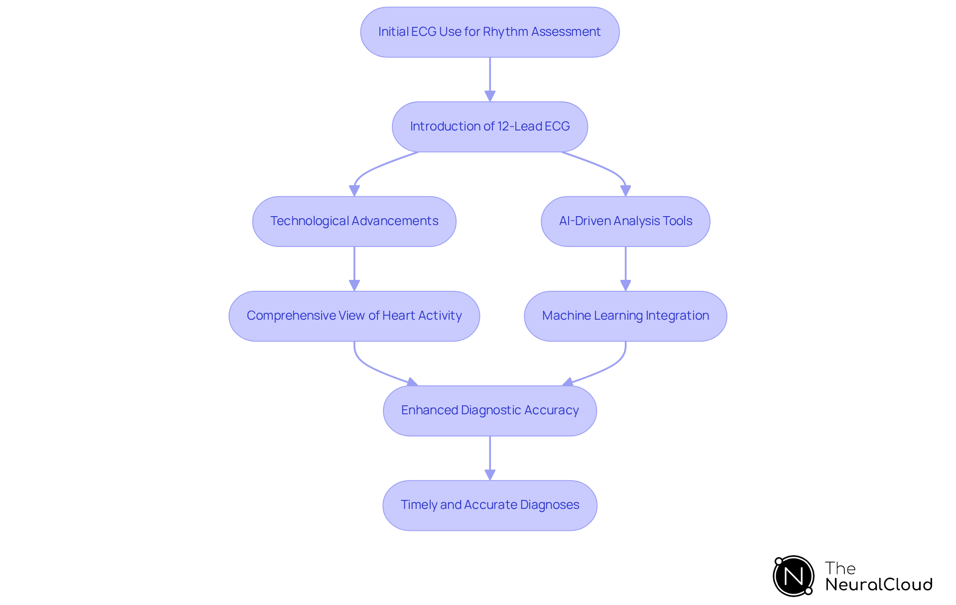
Despite its long history, ECG technology has continually evolved to meet the growing demands of accurate myocardial damage diagnosis. Initially, ECGs were used primarily for basic rhythm assessments. However, as our understanding of cardiac physiology deepened, the ECG has become a vital instrument in accurately diagnosing what does an MI look like on an ECG. The introduction of the 12-lead ECG provided a comprehensive view of the heart's electrical activity, which is essential for understanding what does an MI look like on an ECG and allowing clinicians to pinpoint specific areas of ischemia.
Over the decades, technological advancements, including digital ECG machines and AI-driven analysis tools, have significantly enhanced diagnostic accuracy. Integrating machine learning algorithms into ECG analysis is revolutionizing myocardial infarction detection. These innovations enable quicker and more precise diagnoses, which are essential for improving patient outcomes. Recent studies demonstrate the effectiveness of AI-based ECG interpretation, achieving a sensitivity of 77% and a specificity of 99%. As these technologies advance, they promise to redefine emergency care, making timely and accurate diagnoses a reality for patients.
Discuss the Clinical Implications of MI Detection on ECG
Understanding what does an MI look like on an ECG presents significant clinical challenges that can impact patient outcomes. Quickly identifying MI allows for immediate interventions like thrombolytics or percutaneous coronary interventions. These actions are vital for restoring blood flow and reducing heart damage. Research shows that 25% of individuals classified as 'non-STEMI' actually have acute coronary occlusion (ACO). This misclassification can lead to delayed treatment and increased mortality rates, underscoring the critical role of accurate ECG interpretation in improving patient outcomes.
Understanding what does an MI look like on an ECG, including the specific variations associated with different types of MI such as STEMI and NSTEMI, enables healthcare providers to customize treatment strategies effectively. For instance, ST elevation on an ECG is a key indicator of potential coronary artery occlusion, which can lead to STEMI. The integration of advanced ECG interpretation methods, particularly those enhanced by artificial intelligence, has shown to improve the detection of obstructive MI, achieving an accuracy rate of 84% in clinical settings.
Healthcare professionals need ongoing education and training in ECG interpretation to spot these critical changes and react effectively. The move towards the occlusion MI (OMI) paradigm highlights the importance of a deeper understanding of MI detection. This approach aims to minimize misdiagnoses and improve triage in emergency cardiology. By refining treatment prioritization based on the presence of acute vessel occlusion, healthcare providers can ensure timely and appropriate interventions. Ultimately, refining ECG interpretation can lead to timely interventions that save lives and improve recovery rates.

Conclusion
Recognizing myocardial infarction (MI) on an ECG is essential for timely diagnosis and effective treatment. This article highlights the significance of recognizing key ECG changes, such as:
- ST-segment elevation
- T-wave inversions
- The development of pathological Q waves
These alterations indicate myocardial damage and guide healthcare professionals in making informed clinical decisions that can ultimately save lives.
The discussion emphasizes the importance of advanced technologies, such as Neural Cloud Solutions' MaxYield™, which enhance the accuracy of ECG interpretation. With AI-driven analysis, healthcare providers can achieve higher sensitivity and specificity in identifying obstructive MI, leading to improved patient outcomes. ECG technology has evolved from simple rhythm checks to advanced AI integration, highlighting significant advancements in cardiac diagnostics.
In light of these insights, it is imperative for healthcare professionals to stay informed about the latest developments in ECG interpretation and the clinical implications of MI detection. By embracing innovative tools and refining diagnostic skills, clinicians can ensure timely interventions that significantly improve recovery rates and patient care. Using advanced ECG technologies boosts diagnostic accuracy and is crucial for the future of emergency cardiology, changing how we manage myocardial infarction in clinical practice.
Frequently Asked Questions
What is myocardial infarction (MI)?
Myocardial infarction (MI) occurs when blood flow to a section of the heart is blocked, leading to damage or death of heart muscle tissue, often due to blockages in one or more coronary arteries caused by plaque buildup.
Why is understanding ECG changes important in diagnosing MI?
Understanding ECG changes is crucial because they reveal vital alterations that indicate both the presence and severity of myocardial damage, guiding healthcare professionals in making informed clinical decisions.
What are the key ECG changes that indicate myocardial infarction?
The key ECG changes that indicate myocardial infarction include ST-segment elevation, T-wave inversions, and the development of pathological Q waves.
What is the prevalence of acute myocardial infarction in Canada?
In Canada, acute myocardial infarction remains a significant public health concern, with 51,365 deaths attributed to this condition in the fiscal year 2022-2023.
How can AI methods improve the diagnosis of acute coronary syndrome?
AI methods can enhance the management of suspected acute coronary syndrome by making diagnoses more accurate. For example, a deep learning model achieved an accuracy rate of 84% in identifying obstructive MI, outperforming traditional methods.
What is MaxYield™ and how does it assist in analyzing ECG changes?
MaxYield™ is a product from Neural Cloud Solutions that analyzes ECG changes using advanced noise filtering and automation, transforming noisy recordings into detailed insights. It provides beat-by-beat analysis of 200,000 heartbeats in less than 5 minutes.
How does Insight360 enhance the use of MaxYield™?
Insight360 is a customizable visualization and reporting tool that enhances MaxYield™ by transforming clean ECG data into interactive dashboards and clinical-ready reports, improving diagnostic yield and supporting informed clinical decision-making.
What evidence supports the effectiveness of ECG in diagnosing myocardial infarction?
Case studies have shown that AI models can accurately identify cases of myocardial infarction on initial ECGs, with one study detecting 553 confirmed cases compared to 427 detected by traditional methods, highlighting the critical role of ECG in early detection and intervention.
List of Sources
- Define Myocardial Infarction and Its ECG Significance
- New study finds AI model improves heart attack detection (https://health.ucdavis.edu/news/headlines/new-study-finds-ai-model-improves-heart-attack-detection/2025/11)
- An ECG biomarker for sudden cardiac death discovered with deep learning - Nature (https://nature.com/articles/s41586-026-10674-6)
- AI-based ECG interpretation outperforms standard diagnosis of occlusive myocardial infarction (https://news-medical.net/news/20260323/AI-based-ECG-interpretation-outperforms-standard-diagnosis-of-occlusive-myocardial-infarction.aspx)
- Number of heart disease deaths in Canada 2000-2023| Statista (https://statista.com/statistics/882535/diagnosed-heart-disease-number-of-deaths-canada?srsltid=AfmBOopCjBkZDJizLnbLhOnWC8SeRdOPlxkt1s5hVwwSrnXxGvr0N8Z0)
- Identify Key ECG Features of Myocardial Infarction
- AI-based ECG interpretation outperforms standard diagnosis of occlusive myocardial infarction (https://news-medical.net/news/20260323/AI-based-ECG-interpretation-outperforms-standard-diagnosis-of-occlusive-myocardial-infarction.aspx)
- STEMI to OMI: A Paradigm-Shift Review (https://powerfulmedical.com/research/state-of-the-art-review-from-stsegment-elevation-mi-to-occlusion-mi-the-new-paradigm-shift-in-acute-myocardial-infarction)
- Acute ST-Segment Elevation Myocardial Infarction (STEMI) - StatPearls - NCBI Bookshelf (https://ncbi.nlm.nih.gov/books/NBK532281)
- pmc.ncbi.nlm.nih.gov (https://pmc.ncbi.nlm.nih.gov/articles/PMC10122825)
- ECGs in Acute Myocardial Infarction | ACLS Training (https://aclsmedicaltraining.com/ecg-in-acute-myocardial-infarction)
- Explore the Evolution of MI Diagnosis via ECG
- New study finds AI model improves heart attack detection (https://health.ucdavis.edu/news/headlines/new-study-finds-ai-model-improves-heart-attack-detection/2025/11)
- Artificial intelligence may speed heart attack diagnosis and treatment (https://newsroom.heart.org/news/artificial-intelligence-may-speed-heart-attack-diagnosis-and-treatment)
- AI outperforms conventional diagnosis for certain types of heart attacks: Study (https://health.economictimes.indiatimes.com/amp/news/diagnostics/ai-outperforms-conventional-diagnosis-for-certain-types-of-heart-attacks-study/129746852)
- Frontiers | ECG data analysis to determine ST-segment elevation myocardial infarction and infarction territory type: an integrative approach of artificial intelligence and clinical guidelines (https://frontiersin.org/journals/physiology/articles/10.3389/fphys.2024.1462847/full)
- AI-based ECG interpretation outperforms standard diagnosis of occlusive myocardial infarction (https://news-medical.net/news/20260323/AI-based-ECG-interpretation-outperforms-standard-diagnosis-of-occlusive-myocardial-infarction.aspx)
- Discuss the Clinical Implications of MI Detection on ECG
- AI-based ECG interpretation outperforms standard diagnosis of occlusive myocardial infarction (https://news-medical.net/news/20260323/AI-based-ECG-interpretation-outperforms-standard-diagnosis-of-occlusive-myocardial-infarction.aspx)
- AI ECG Better Detects Severe Heart Attacks in Emergency Setting - American College of Cardiology (https://acc.org/about-acc/press-releases/2025/10/28/18/55/ai-ecg-better-detects-severe-heart-attacks-in-emergency-setting)
- STEMI to OMI: A Paradigm-Shift Review (https://powerfulmedical.com/research/state-of-the-art-review-from-stsegment-elevation-mi-to-occlusion-mi-the-new-paradigm-shift-in-acute-myocardial-infarction)