Introduction
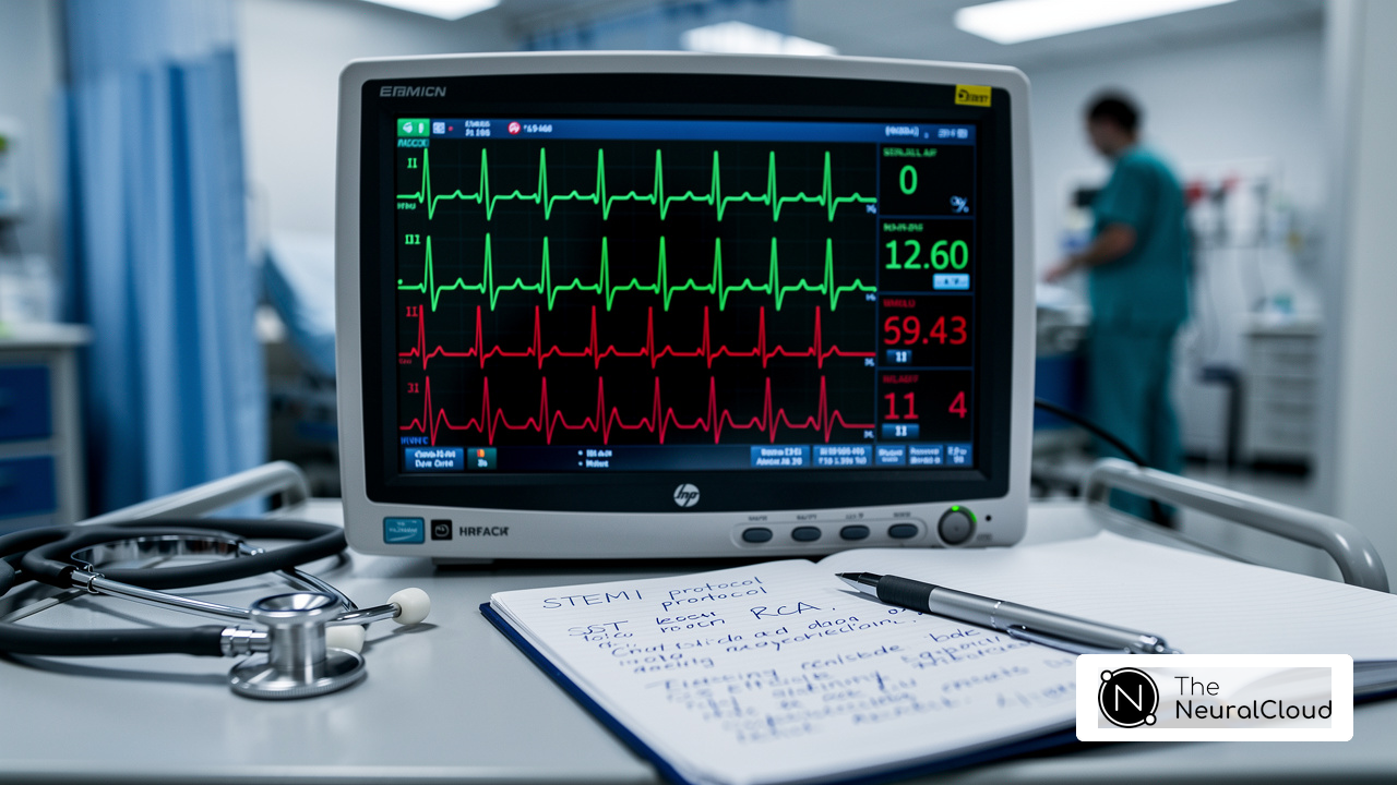
Clinicians face significant challenges in accurately identifying idioventricular rhythm (IVR) during cardiac monitoring. IVR is marked by wide QRS complexes and a lack of normal atrial activity, often appearing in critical conditions like myocardial ischemia. By understanding IVR's ECG presentation and clinical significance, clinicians can make informed treatment decisions that positively impact patient outcomes.
Identifying and managing IVR presents challenges, but advanced technologies like MaxYield can enhance diagnostic confidence.
Define Idioventricular Rhythm
Idioventricular activity (IVR) presents unique challenges in cardiac monitoring, particularly due to its atypical impulse generation. IVR is characterized by the absence of normal atrial function, where the heart's lower chambers, or ventricles, produce impulses at a rate typically below 50 beats per minute. This pattern occurs when ectopic pacemakers in the ventricles take over, often due to failures in the higher pacemaker sites like the sinoatrial (SA) node or atrioventricular (AV) node.
Clinically, when asking what does idioventricular rhythm look like on an electrocardiogram (ECG), it is identifiable as a consistent pattern with wide QRS complexes, indicating that impulses originate from the ventricles rather than the atria. Most cases of IVR are benign and self-limiting, resolving without intervention. However, persistent symptoms such as weariness, lightheadedness, fainting, and heart palpitations can complicate patient care, requiring timely medical evaluation.
It's crucial for healthcare professionals to grasp this definition to effectively diagnose and manage cardiac conditions. Additionally, accelerated idioventricular rhythm (AIVR), a variant with a heart rate between 50 and 110 beats per minute, can occur in specific clinical scenarios, such as during myocardial infarction. This highlights the importance of vigilant monitoring and intervention in patients with AIVR.
With MaxYield, a device-agnostic ECG intelligence layer, healthcare professionals can utilize advanced ECG analysis. This ensures clean data and hospital-grade accuracy, significantly enhancing monitoring capabilities. Expert endorsements from Dr. Alan Rabinowitz, Dr. Brett Heilbron, and Dr. Marc W. Deyell emphasize that MaxYield’s precision rivals human interpretation, reducing noise and improving diagnostic confidence. Identifying the underlying causes and monitoring for symptoms of IVR is essential for effective care, particularly when leveraging MaxYield’s capabilities, which seamlessly integrate with existing systems to offer actionable insights.

Context and Importance of Idioventricular Rhythm
Idioventricular rhythm (IVR) presents a significant challenge in clinical settings, particularly during myocardial ischemia or after reperfusion therapy in acute myocardial infarction cases. Its detection can indicate underlying cardiac issues, such as structural heart disease or electrolyte disturbances. Spotting IVR is crucial because it indicates how severe the cardiac condition might be and can directly impact treatment choices. For instance, in individuals with acute coronary syndromes, identifying IVR may lead to further diagnostic evaluations and interventions aimed at reducing potential complications.
Additionally, understanding the context of IVR is vital for distinguishing it from other arrhythmias, like ventricular tachycardia, which may require more aggressive management. Research shows a strong link between IVR and myocardial ischemia-reperfusion, highlighting its role as a key indicator of myocardial necrosis. In a study involving 87 individuals with persistent ischemic chest pain, IVR was identified as a strong predictor of both myocardial necrosis and reperfusion, underscoring its clinical significance.
Furthermore, the presence of IVR can guide healthcare professionals in making informed decisions regarding patient care. This recognition can lead to tailored treatment plans that address the underlying cardiac issues more effectively, ultimately enhancing outcomes in cardiovascular management.

Characteristics and ECG Presentation of Idioventricular Rhythm
Interpreting ECGs can be challenging, especially when noise and artifact obscure critical data. The ECG display of idioventricular activity (IVR) provides insight into what does idioventricular rhythm look like, characterized by a consistent ventricular pattern, typically with a rate under 50 beats per minute. When considering what does idioventricular rhythm look like, key features include the absence of P waves, indicating a lack of atrial activity, and a widened QRS complex, generally exceeding 120 milliseconds. The pattern often shows a consistent R-R interval, reflecting the regularity of ventricular contractions. It is essential for healthcare technology developers to understand what does idioventricular rhythm look like, as the idioventricular pattern is usually harmless and often resolves without treatment.
In instances of accelerated idioventricular activity (AIVR), the ventricular rate can vary from 50 to 120 beats per minute, while the QRS complexes remain wide. Recognizing these ECG features is crucial for clinicians, as they aid in differentiating IVR from other arrhythmias, such as ventricular tachycardia, which may require distinct management strategies. AIVR is often associated with increased vagal tone and can occur post-myocardial infarction during the reperfusion phase, highlighting the importance of context in ECG interpretation.
Neural Cloud Solutions Inc.'s MaxYield™ platform plays a pivotal role in enhancing the analysis of these patterns. MaxYield™ uses advanced noise filtering and wave recognition to isolate and label critical data, even in noisy recordings. This helps clinicians interpret ECGs with confidence, especially in complex cases like AIVR, where precise identification of features is crucial for effective management. Expert endorsements from Dr. Alan Rabinowitz, Dr. Brett Heilbron, and Dr. Marc W. Deyell emphasize that MaxYield™'s precision rivals human interpretation, significantly improving diagnostic yield.
To reduce the risk of idioventricular rhythm, consider these practices:
- Maintain normal electrolyte levels
- Engage in heart-healthy practices
Case studies demonstrate that AIVR can occur in individuals without underlying cardiac disease, highlighting the necessity for careful monitoring and evaluation of individual circumstances. For instance, AIVR may occur following the return of spontaneous circulation after cardiac arrest, likely related to the reperfusion of the heart. Understanding these connections is vital for managing patients effectively, and integrating MaxYield™ into clinical workflows not only simplifies ECG analysis but also enhances patient care outcomes.

Causes and Clinical Implications of Idioventricular Rhythm
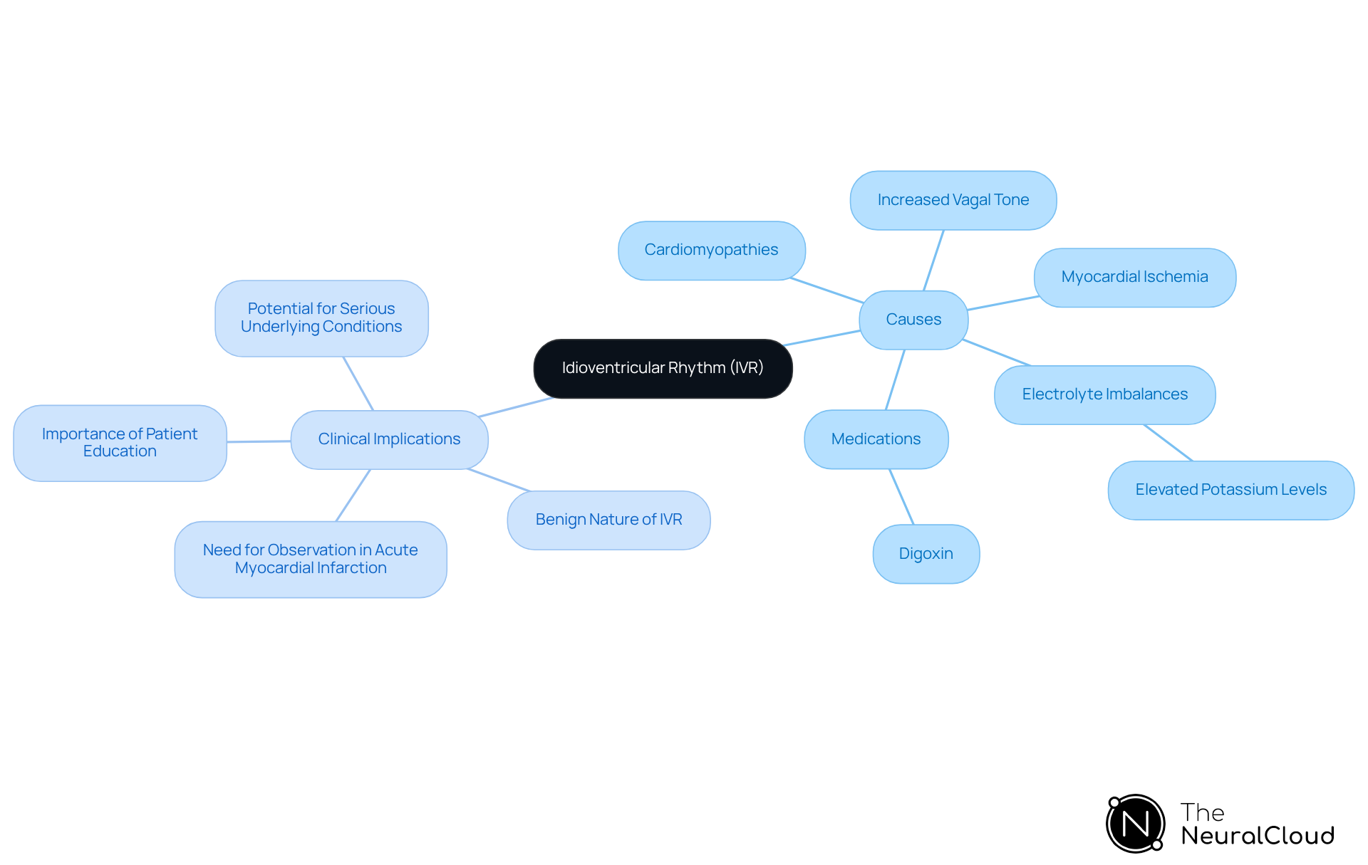
Idioventricular activity (IVR) presents a unique challenge in ECG analysis, stemming from various underlying causes, including:
- Myocardial ischemia
- Cardiomyopathies
- Electrolyte imbalances, particularly elevated potassium levels
It may also arise from increased vagal tone or certain medications, such as digoxin. Clinically, IVR is usually seen as benign and self-limiting, especially in the absence of structural heart disease. However, recognizing IVR is crucial, as it may signal serious underlying conditions needing attention. For instance, in individuals with acute myocardial infarction, the emergence of IVR may indicate a need for closer observation and possible therapeutic actions to address the underlying ischemia.
A small study found that accelerated idioventricular activity (AIVR) was associated with a lower 7-day survival rate in post-resuscitation patients, highlighting the importance of understanding these patterns in clinical settings. Moreover, educating and reassuring patients is essential, as many instances of IVR are self-limiting and resolve when sinus frequency surpasses that of ventricular foci. Understanding IVR is not just about recognizing a rhythm; it’s about ensuring that patients receive the appropriate care they need.
Conclusion
Understanding idioventricular rhythm (IVR) is vital for effective cardiac monitoring and patient management. Recognizing IVR plays a significant role in identifying potential underlying cardiac issues and informs treatment decisions that can greatly impact patient outcomes.
Key insights discussed include the distinctive features of IVR on an ECG, such as the absence of P waves and wide QRS complexes. These characteristics help differentiate IVR from other arrhythmias, particularly in contexts like myocardial ischemia, where IVR may indicate serious underlying conditions.
Furthermore, the integration of advanced ECG analysis tools like MaxYield™ enhances the ability to monitor and interpret these rhythms accurately. This ensures that clinicians can provide timely and effective care, ultimately improving patient outcomes.
In conclusion, recognizing and understanding idioventricular rhythm is essential for clinicians and diagnostic facilities alike. By leveraging advanced technologies and maintaining a keen awareness of the clinical implications of IVR, healthcare professionals can significantly enhance patient safety and care quality.
Frequently Asked Questions
What is idioventricular rhythm (IVR)?
Idioventricular rhythm (IVR) is characterized by the heart's lower chambers, or ventricles, generating impulses at a rate typically below 50 beats per minute, due to the absence of normal atrial function. This occurs when ectopic pacemakers in the ventricles take over, often due to failures in higher pacemaker sites like the sinoatrial (SA) node or atrioventricular (AV) node.
How can idioventricular rhythm be identified on an electrocardiogram (ECG)?
IVR can be identified on an ECG by a consistent pattern with wide QRS complexes, indicating that the impulses originate from the ventricles rather than the atria.
Are there any symptoms associated with idioventricular rhythm?
While most cases of IVR are benign and self-limiting, persistent symptoms such as weariness, lightheadedness, fainting, and heart palpitations can complicate patient care and may require timely medical evaluation.
What is accelerated idioventricular rhythm (AIVR)?
Accelerated idioventricular rhythm (AIVR) is a variant of IVR characterized by a heart rate between 50 and 110 beats per minute, which can occur in specific clinical scenarios, such as during myocardial infarction.
Why is it important for healthcare professionals to understand idioventricular rhythm?
Understanding IVR is crucial for healthcare professionals to effectively diagnose and manage cardiac conditions, particularly in recognizing symptoms and determining the need for intervention.
How does MaxYield enhance the monitoring of idioventricular rhythm?
MaxYield, a device-agnostic ECG intelligence layer, provides advanced ECG analysis that ensures clean data and hospital-grade accuracy, significantly enhancing monitoring capabilities and diagnostic confidence.
What do expert endorsements say about MaxYield's capabilities?
Experts such as Dr. Alan Rabinowitz, Dr. Brett Heilbron, and Dr. Marc W. Deyell endorse MaxYield, stating that its precision rivals human interpretation, reducing noise and improving diagnostic confidence in cardiac monitoring.
List of Sources
- Define Idioventricular Rhythm
- Idioventricular Rhythm - StatPearls - NCBI Bookshelf (https://ncbi.nlm.nih.gov/books/NBK554520)
- What Is Idioventricular Rhythm? (https://icliniq.com/articles/heart-circulatory-health/idioventricular-rhythm)
- Idioventricular Rhythm: Definition, Symptoms and Treatment (https://my.clevelandclinic.org/health/diseases/23070-idioventricular-rhythm)
- Context and Importance of Idioventricular Rhythm
- Usefulness of the accelerated idioventricular rhythm as a marker for myocardial necrosis and reperfusion during thrombolytic therapy in acute myocardial infarction (https://research.rug.nl/en/publications/usefulness-of-the-accelerated-idioventricular-rhythm-as-a-marker-)
- Ventricular Rhythms with Intermediate Rates in Acute Myocardial Infarction (https://sciencedirect.com/science/article/abs/pii/S0012369216395113)
- Accelerated Idioventricular Rhythm: Background, Pathophysiology, Etiology (https://emedicine.medscape.com/article/150074-overview)
- Browse issues - Journal of Surgery and Trauma (https://jsurgery.bums.ac.ir/browse.php?a_id=477&sid=1&slc_lang=en&ftxt=1)
- Prevalence and significance of accelerated idioventricular rhythm in patients with ST-elevation myocardial infarction treated with primary percutaneous coronary intervention - PubMed (https://pubmed.ncbi.nlm.nih.gov/19962468)
- Characteristics and ECG Presentation of Idioventricular Rhythm
- Idioventricular Rhythm: Definition, Symptoms and Treatment (https://my.clevelandclinic.org/health/diseases/23070-idioventricular-rhythm)
- Idioventricular Rhythm - StatPearls - NCBI Bookshelf (https://ncbi.nlm.nih.gov/books/NBK554520)
- Accelerated Idioventricular Rhythm (AIVR) (https://litfl.com/accelerated-idioventricular-rhythm-aivr)
- Causes and Clinical Implications of Idioventricular Rhythm
- Idioventricular Rhythm - StatPearls - NCBI Bookshelf (https://ncbi.nlm.nih.gov/books/NBK554520)
- Accelerated Idioventricular Rhythm: Background, Pathophysiology, Etiology (https://emedicine.medscape.com/article/150074-overview)