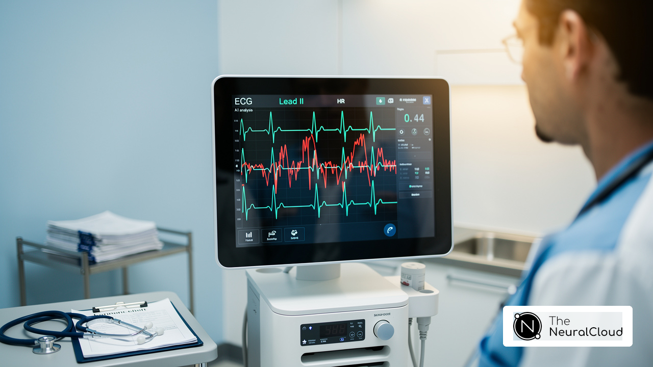
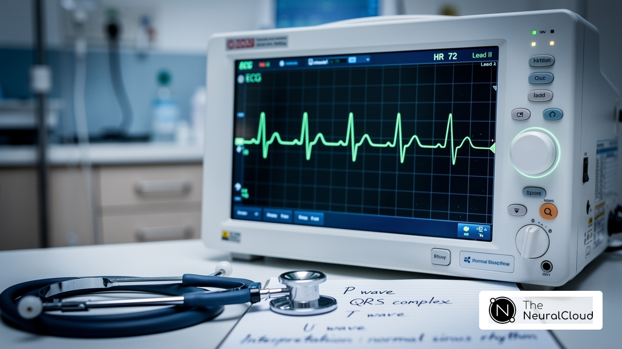
Introduction
Hypokalemia, a condition marked by dangerously low potassium levels, poses significant risks to cardiac health, particularly in vulnerable populations such as older adults and those with heart conditions. Recognizing EKG changes linked to hypokalemia is essential for healthcare professionals because these changes can indicate serious complications, such as arrhythmias. Yet, accurately spotting these subtle changes on an electrocardiogram can be challenging.
How can clinicians effectively interpret EKG results to ensure timely intervention and improve patient outcomes in the face of this potentially life-threatening condition?
Define Hypokalemia and Its Cardiac Implications
Hypokalemia, characterized by serum potassium levels below 3.5 mmol/L, poses significant risks to cardiac health, particularly in vulnerable populations. Potassium is vital for normal cardiac function, influencing the heart's electrical activity. Low levels of this mineral can lead to severe complications, such as arrhythmias, muscle weakness, and potentially cardiac arrest. The heart's electrical conduction system relies on potassium to maintain the resting membrane potential and facilitate repolarization. When potassium levels decrease, this balance is disrupted, leading to EKG changes with hypokalemia.
In Canada, studies indicate that 19% to 54% of patients receiving treatment for heart failure may experience low potassium levels, often exacerbated by diuretics. This underscores why monitoring potassium levels is so critical, especially for older adults and those with existing heart conditions who face a higher risk of arrhythmias. Expert opinions emphasize that prompt diagnosis and intervention are essential, as untreated low potassium levels can lead to life-threatening cardiac events. For healthcare professionals, recognizing and addressing hypokalemia is crucial to preventing potentially fatal cardiac events in at-risk patients.

Identify ECG Changes Linked to Hypokalemia
Recognizing EKG changes with hypokalemia is vital for timely diagnosis and effective treatment. Key alterations include:
- T-wave flattening or inversion: Often one of the earliest indicators, this change signifies altered repolarization and can be a precursor to more severe complications.
- ST-segment depression: This reflects impaired myocardial repolarization and may suggest ischemia, highlighting the need for careful monitoring.
- Prominent U waves: These waves, which appear following the T wave, are a hallmark of low potassium levels and are particularly prevalent in severe cases, serving as a reliable marker for clinicians.
- Prolonged QT interval: This alteration increases the risk of life-threatening arrhythmias, necessitating prompt intervention.
- Increased amplitude of P waves: This modification may indicate atrial enlargement due to altered electrical conduction, further complicating the clinical picture.
It's essential for healthcare professionals to spot EKG changes with hypokalemia to diagnose low potassium levels accurately and choose the right treatment, ultimately improving patient outcomes. As highlighted by specialist Srinivasan Ramadurai, 'EKG changes with hypokalemia, particularly the presence of U waves, ST depression, T-wave flattening/inversion, and QT prolongation, are reliable indicators of the severity of potassium deficiency, supporting the use of EKG as a rapid bedside diagnostic tool and guiding urgent management.' Furthermore, low potassium levels are categorized into mild (3.0 - 3.4 mmol/L), moderate (2.5 - 2.9 mmol/L), and severe (≤ 2.4 mmol/L), which aids in understanding the EKG changes with hypokalemia in relation to the severity of the condition. This underscores the importance of integrating advanced tools like MaxYield™ to enhance diagnostic accuracy and patient care in clinical practice. MaxYield™ from Neural Cloud Solutions Inc. can enhance this process by automating ECG analysis, reducing noise, and providing healthcare professionals with clear, actionable insights to support clinical decision-making. MaxYield™ is currently pending FDA Class II SaMD clearance, ensuring compliance with regulatory standards and enhancing its credibility in clinical settings.

Implement Advanced ECG Analysis Techniques
Accurate ECG interpretation in hypokalemia cases can be challenging due to subtle EKG changes with hypokalemia that are often overlooked. To enhance the accuracy of ECG interpretation, consider implementing the following advanced techniques:
- Machine Learning Algorithms: Leverage AI-driven tools like MaxYield™ that analyze ECG patterns to detect subtle changes associated with hypokalemia. Recent advancements in deep learning have shown potential in enhancing the sensitivity of ECG for identifying elevated electrolyte levels, with models achieving an area under the curve (AUC) of 0.945 for hyperkalemia detection. Experts such as Dr. Alan Rabinowitz emphasize that MaxYield™ rivals human interpretation in precision, making it a valuable asset in clinical settings.
- Signal Processing Techniques: Employ advanced filtering methods through MaxYield™ to isolate noise and enhance the clarity of ECG signals. This is essential for improving the visibility of T waves and U waves, which are frequently influenced by imbalances in mineral levels, such as EKG changes with hypokalemia. The absence of P waves signifies severely elevated potassium levels, making clear signal processing essential. MaxYield™ maps ECG signals through noise, isolating and labeling key features in every heartbeat, thus transforming noisy recordings into detailed insights.
- Automated Reporting Tools: Utilize Insight360, the customizable visualization tool that generates reports highlighting key ECG changes, facilitating quicker clinical decision-making. Automated tools can streamline workflows by ensuring that healthcare providers can promptly identify significant ECG alterations, such as peaked T waves and prolonged PR intervals, which are indicative of hyperkalemia. With MaxYield™, you get beat-by-beat analysis that processes 200,000 heartbeats in under 5 minutes, helping you make confident clinical decisions.
- Integration with Clinical Data: Combine ECG findings with patient history and laboratory results to provide a comprehensive view of the patient's condition. This holistic approach allows for proactive monitoring and swift clinical responses, which are vital for improving patient safety and outcomes in cases of electrolyte imbalances. With MaxYield™, healthcare providers can scale clean signals and clear data across their entire workflow, ensuring audit-ready documentation supporting GxP inspections.
These techniques not only enhance diagnostic accuracy but also streamline workflows for healthcare providers, ultimately leading to better patient care. By adopting these advanced techniques, healthcare providers can enhance patient care and ensure timely clinical responses to electrolyte imbalances. For those interested, Neural Cloud Solutions Inc. offers free trials and personalized walkthroughs of MaxYield™ and Insight360.

Analyze and Interpret ECG Results in Hypokalemia
Analyzing EKG changes with hypokalemia presents unique challenges that require careful attention. Here’s a structured approach to ensure accurate interpretation and effective management:
- Review the Patient's History: Start by assessing symptoms such as muscle weakness, palpitations, or arrhythmias that may correlate with ECG findings. Low potassium levels can lead to serious complications, including life-threatening arrhythmias, making it essential to monitor EKG changes with hypokalemia in patient history.
- Identify Key ECG Variations: Look for characteristic alterations such as T-wave flattening, ST-segment depression, prominent U waves, and prolonged QT intervals. These alterations serve as vital markers for clinicians, emphasizing the need for continuous monitoring and timely intervention.
- Assess Severity: Evaluate the seriousness of hypokalemia based on the observed ECG variations and correlate them with serum electrolyte levels. You can identify hypokalemia when potassium levels drop below 3.5 mmol/L, with severe instances dropping below 2.5 mmol/L, which can lead to EKG changes with hypokalemia.
- Consider Differential Diagnoses: Rule out other causes of similar ECG variations, such as ischemia or other electrolyte imbalances. Understanding the electrophysiological mechanisms underlying these changes is essential for accurate diagnosis and effective management.
- Plan Management: Based on the findings, decide on the appropriate management strategy, which may include mineral supplementation or further cardiac monitoring. Effective management strategies for low potassium levels involve oral chloride for mild cases and intravenous solutions for severe cases, alongside dietary modifications.
By carefully analyzing ECG results, healthcare providers can swiftly manage low potassium levels, ensuring better patient outcomes. Recent studies indicate that dietary interventions can significantly lower cardiovascular risks associated with hypokalemia, highlighting the importance of monitoring potassium levels, especially in patients with pre-existing heart conditions.

Conclusion
Hypokalemia poses significant challenges for healthcare professionals, particularly in recognizing its impact on cardiac health. This tutorial has highlighted the significant risks associated with low potassium levels, particularly how they manifest in EKG changes. By recognizing these alterations, clinicians can take timely action to prevent severe complications, ensuring better patient outcomes.
Key insights discussed include:
- Identification of specific EKG changes:
- T-wave flattening
- ST-segment depression
- Prominent U waves
These serve as vital indicators of hypokalemia. The tutorial also emphasized the importance of advanced ECG analysis techniques, including the use of tools like MaxYield™ from Neural Cloud Solutions Inc., which can enhance diagnostic accuracy and streamline clinical workflows. This integration not only enhances diagnostic accuracy but also empowers clinicians to provide timely interventions.
By prioritizing the understanding of EKG changes associated with hypokalemia, clinicians can safeguard their patients against potentially fatal cardiac events. As the prevalence of low potassium levels continues to pose risks, especially among vulnerable populations in Canada, it is imperative for clinicians to remain vigilant. By adopting a proactive approach to monitoring and interpreting ECG results, healthcare professionals can significantly reduce the risk of life-threatening cardiac events, ultimately enhancing patient safety and care.
Frequently Asked Questions
What is hypokalemia?
Hypokalemia is defined as having serum potassium levels below 3.5 mmol/L, which can pose significant risks to cardiac health.
Why is potassium important for cardiac function?
Potassium is vital for normal cardiac function as it influences the heart's electrical activity, helping to maintain the resting membrane potential and facilitate repolarization.
What are the potential complications of hypokalemia?
Complications of hypokalemia can include arrhythmias, muscle weakness, and potentially cardiac arrest.
How does hypokalemia affect the heart's electrical conduction system?
Low potassium levels disrupt the balance necessary for the heart's electrical conduction system, leading to EKG changes associated with hypokalemia.
What percentage of heart failure patients in Canada may experience low potassium levels?
Studies in Canada indicate that between 19% to 54% of patients receiving treatment for heart failure may experience low potassium levels, often exacerbated by diuretics.
Who is at higher risk for hypokalemia-related complications?
Older adults and individuals with existing heart conditions are at a higher risk of experiencing arrhythmias due to low potassium levels.
Why is monitoring potassium levels critical in certain populations?
Monitoring potassium levels is critical, especially for vulnerable populations like older adults and those with heart conditions, to prevent potentially life-threatening cardiac events.
What is the importance of prompt diagnosis and intervention for hypokalemia?
Prompt diagnosis and intervention are essential because untreated low potassium levels can lead to serious and life-threatening cardiac events.
List of Sources
- Define Hypokalemia and Its Cardiac Implications
- Hypokalemia: Background, Etiology, Pathophysiology (https://emedicine.medscape.com/article/242008-overview)
- Hypokalemia and arrhythmia: Is there a link? (https://medicalnewstoday.com/articles/hypokalemia-arrhythmia)
- Hypokalemia-Induced Life-Threatening Arrhythmia in a Patient With Congestive Heart Failure (https://cureus.com/articles/135236-hypokalemia-induced-life-threatening-arrhythmia-in-a-patient-with-congestive-heart-failure)
- Boosting Potassium Helps Patients at High Risk for Ventricular Arrhythmias (https://tctmd.com/news/boosting-potassium-helps-patients-high-risk-ventricular-arrhythmias)
- Identify ECG Changes Linked to Hypokalemia
- Hypokalaemia (https://litfl.com/hypokalaemia-ecg-library)
- Electrocardiographic Changes in Patients With Hypokalemia and Their Correlation With Serum Potassium Levels - PMC (https://pmc.ncbi.nlm.nih.gov/articles/PMC12510615)
- Electrocardiographic manifestations in severe hypokalemia - PMC (https://pmc.ncbi.nlm.nih.gov/articles/PMC7287199)
- Electrocardiographic Changes in Patients With Hypokalemia and Their Correlation With Serum Potassium Levels (https://cureus.com/articles/400549-electrocardiographic-changes-in-patients-with-hypokalemia-and-their-correlation-with-serum-potassium-levels)
- Implement Advanced ECG Analysis Techniques
- AI ECG Alerts Improve Potassium Imbalance Treatment (https://scienmag.com/ai-ecg-alerts-improve-potassium-imbalance-treatment)
- theneuralcloud.com (https://theneuralcloud.com/post/master-ecg-for-hyperkalemia-key-changes-and-analysis-techniques)
- Deep Learning Model Detects Electrolyte Imbalance via ECG | Docwire News (https://docwirenews.com/post/deep-learning-model-electrolyte-imbalance-ecg)
- Noninvasive Hypokalemia Detection from Single-Lead AI-ECG: Development, Multicenter Validation, and Prospective Pilot Study in the Emergency Department (https://medrxiv.org/content/10.64898/2026.05.23.26353774v1)
- Analyze and Interpret ECG Results in Hypokalemia
- Electrocardiographic manifestations in severe hypokalemia - PMC (https://pmc.ncbi.nlm.nih.gov/articles/PMC7287199)
- theneuralcloud.com (https://theneuralcloud.com/post/understanding-hypokalemia-ecg-changes-for-better-diagnosis)
- theneuralcloud.com (https://theneuralcloud.com/post/understanding-low-potassium-ecg-changes-key-insights-for-developers)
- Hypokalaemia (https://litfl.com/hypokalaemia-ecg-library)