Overview
The article addresses the essential components of normal ECG waves, particularly aimed at developers who require a solid understanding for effective ECG interpretation. It highlights the importance of comprehending elements such as the P-wave, QRS complex, ST segment, and T-wave for diagnosing cardiac conditions. Furthermore, it explores how advanced technologies, notably the MaxYield™ platform, significantly enhance the accuracy and efficiency of ECG analysis. By employing AI and signal processing techniques, the platform provides healthcare professionals with improved diagnostic capabilities.
In the realm of ECG analysis, professionals often face challenges related to the interpretation of complex waveforms. The MaxYield™ platform stands out by offering features that streamline this process. Its integration of AI allows for real-time analysis, reducing the time needed for interpretation while increasing reliability. This capability not only aids in swift decision-making but also minimizes the risk of human error, which is crucial in cardiac care.
The advantages of utilizing the MaxYield™ platform are clear. Healthcare professionals can expect enhanced accuracy in readings, leading to more informed treatment decisions. The platform's user-friendly interface ensures that both technical and non-technical users can navigate its features effectively. As a result, it fosters a collaborative environment where all team members can contribute to patient care with confidence.
In summary, understanding the critical components of ECG waves and leveraging advanced tools like the MaxYield™ platform can transform ECG analysis. By addressing the inherent challenges and providing robust solutions, healthcare professionals are better equipped to diagnose and manage cardiac conditions effectively.
Introduction
Understanding the intricacies of electrocardiograms (ECGs) is crucial for developers focused on enhancing cardiac health monitoring tools. The ECG waveform comprises several key components, including:
- P-wave
- QRS complex
- ST segment
- T-wave
Each playing an essential role in diagnosing heart conditions. Effective clinical decision-making relies on accurately interpreting these signals; however, challenges arise due to noise and artifacts. Developers must explore advanced technologies to improve the precision of ECG analysis, ultimately leading to better patient outcomes.
Define ECG Waves: Key Components and Their Significance
Electrocardiograms (ECGs) provide a graphical representation of the heart's electrical activity, consisting of several critical components:
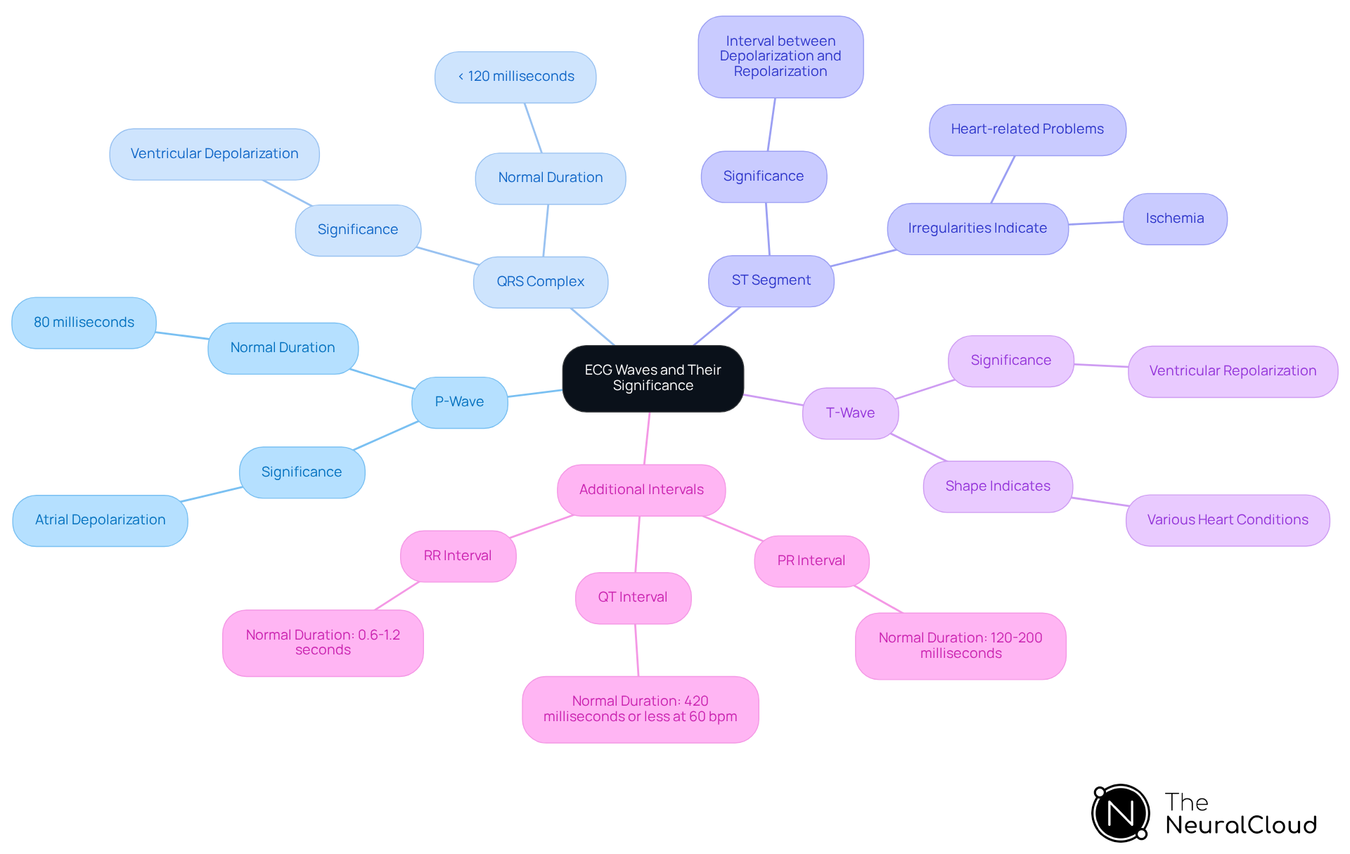
- P-Wave: This wave signifies atrial depolarization, marking the electrical impulse that initiates atrial contraction. A normal P wave duration is approximately 80 milliseconds, which is essential for assessing atrial function.
- QRS Complex: Comprising three distinct waves (Q, R, and S), the QRS complex represents ventricular depolarization, a crucial process for effective heart pumping. The duration of this complex is vital for evaluating ventricular function and should typically be less than 120 milliseconds.
- ST Segment: This segment indicates the interval between ventricular depolarization and repolarization, offering insights into the heart's recovery phase. Irregularities in the ST segment can indicate ischemia or other heart-related problems.
- T-Wave: Representing ventricular repolarization, the T-wave illustrates the heart's return to its resting state. Comprehending the T-wave is essential, as its shape can signify various heart conditions.
Alongside these elements, the PR interval (120-200 milliseconds) and QT interval (420 milliseconds or less at 60 bpm) are essential for understanding conduction durations and identifying irregularities in heart conduction and function. Each component is crucial to diagnosing cardiac conditions, making it essential for developers to understand their importance in recognizing when ECG waves are normal during ECG interpretation.
The MaxYield™ platform from Neural Cloud Solutions enhances this process by utilizing advanced noise filtering and wave recognition techniques. This enables the even amid artifacts. MaxYield™ can recover previously hidden parts of extensive Holter, 1-Lead, and patch monitor recordings, transforming noisy recordings into detailed insights. It provides beat-by-beat evaluations of up to 200,000 heartbeats in under 5 minutes. This capability not only improves diagnostic accuracy but also supports confident clinical decision-making.
Lawrence Rosenthal, MD, PhD, emphasizes that "Tachycardias generated from abnormal repolarization result from genetic defects in ion channels (so-called channelopathies) and can be lethal." By understanding these key components and their implications, developers can leverage the capabilities of MaxYield™ to contribute to more effective ECG analysis tools, ultimately improving patient outcomes.
Analyze the P-Wave, QRS Complex, ST Segment, and T-Wave
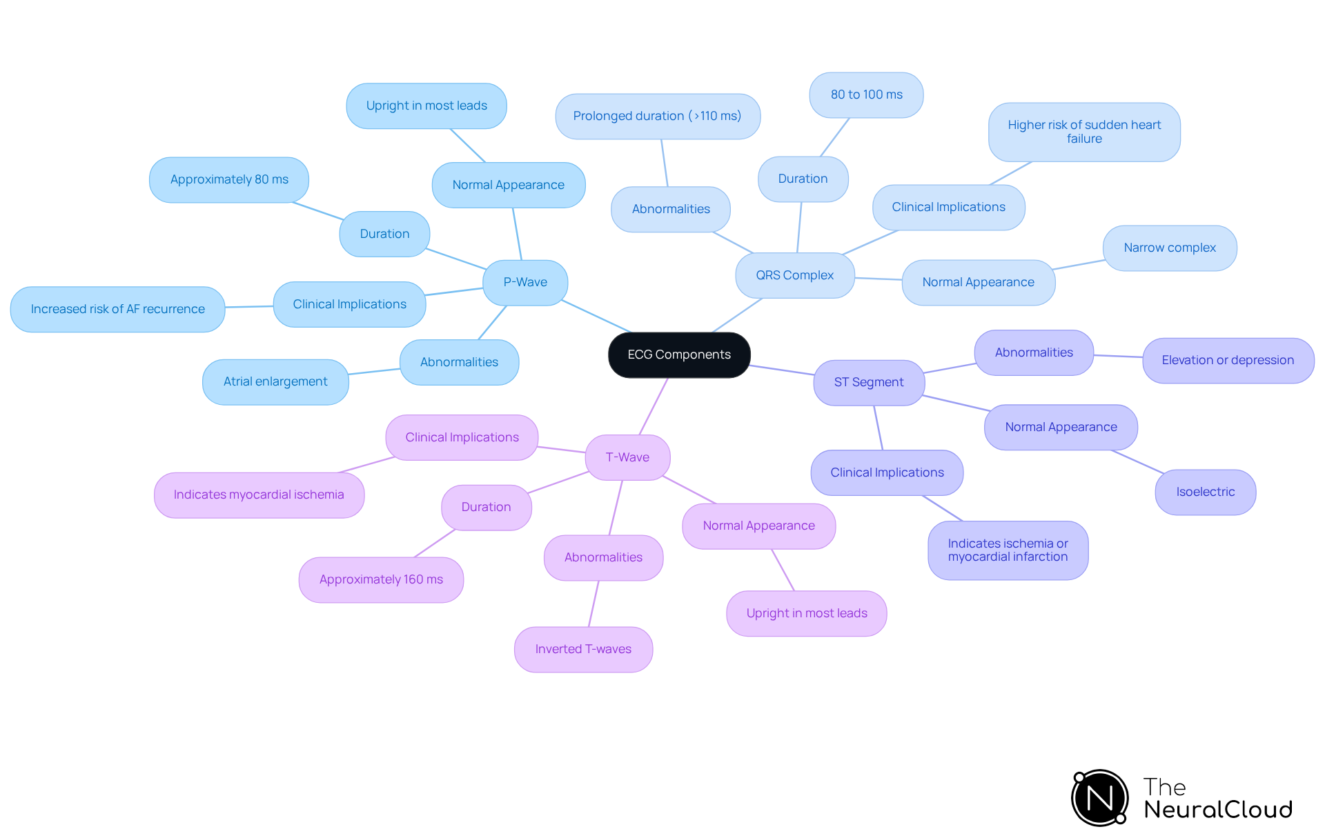
- P-Wave: The P-wave typically lasts about 80 milliseconds and is usually upright in most leads. Abnormalities in the P-wave can indicate conditions such as atrial enlargement. This is clinically significant as it may correlate with increased risks of atrial fibrillation (AF) recurrence after procedures like radiofrequency ablation.
- QRS Complex: Lasting between 80 to 100 milliseconds, a normal QRS complex is narrow, indicating efficient ventricular depolarization. Variations in the , especially when extended beyond 110 milliseconds, can indicate bundle branch blocks or other conduction irregularities. These variations are essential for diagnosing heart problems. For example, a prolonged QRS duration has been linked to higher risks of sudden heart failure.
- The ST segment should be isoelectric in ECG waves normal. Deviations from this baseline, such as elevation or depression, can indicate ischemia or myocardial infarction. This makes it essential for developers to incorporate these parameters into diagnostic algorithms.
- T-Wave: Normally upright in most leads, the T-wave has a duration of about 160 milliseconds. Inverted T-waves may indicate myocardial ischemia or other heart-related issues. This emphasizes the significance of precise T-wave evaluation in clinical environments.
Comprehending these elements and their clinical significance enables developers to improve their algorithms. This enhancement leads to increased diagnostic precision and ultimately benefits health outcomes.
Implement Effective ECG Interpretation Techniques for Clinical Practice
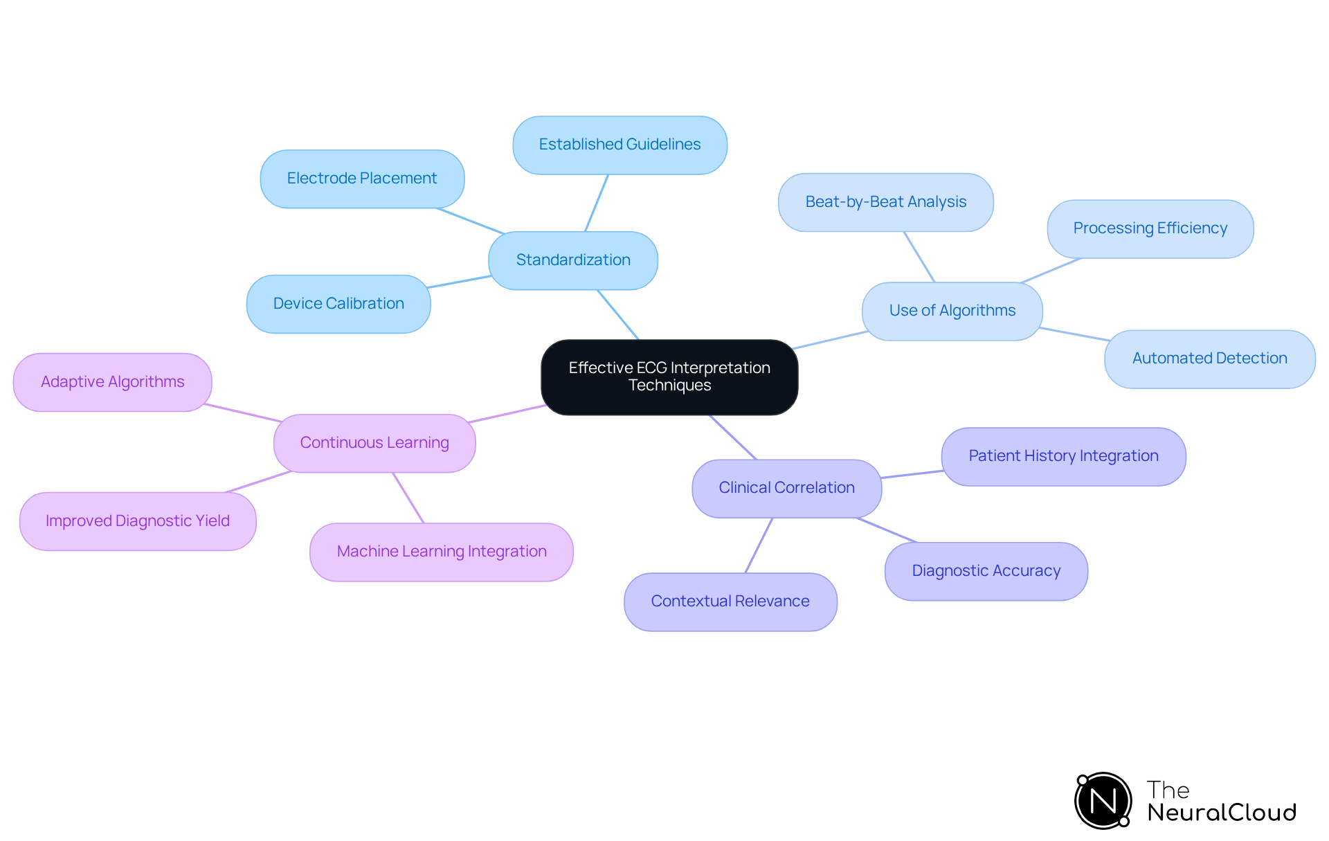
- Standardization: Standardizing ECG recordings is essential for minimizing variability and ensuring accuracy. This process requires meticulous electrode placement and device calibration, following established guidelines to maintain consistency across recordings. By adhering to these protocols, healthcare professionals can enhance the reliability of ECG data.
- Use of Algorithms: The implementation of automated algorithms significantly boosts the efficiency of ECG interpretation. These algorithms accurately detect and label key components of the ECG waves normal, including the P-wave, QRS complex, and T-wave. For example, advanced algorithms can process over 200,000 heartbeats in under five minutes, providing beat-by-beat analysis that isolates and identifies critical features of the ECG waveform, thereby facilitating timely decision-making.
- Clinical Correlation: Correlating ECG findings with clinical symptoms and medical history is crucial. This holistic approach not only enhances diagnostic accuracy but also ensures that the interpretation of ECG data remains contextually relevant. By considering comprehensive patient information, healthcare professionals can make informed decisions that improve patient care.
- Continuous Learning: Employing machine learning methods enables ECG evaluation systems to learn continuously from new data. This adaptive capability improves the interpretation of ECG signals over time, leading to in diagnosing cardiac conditions. The integration of continuous learning models ensures that algorithms evolve, maximizing diagnostic yield and optimizing resource allocation.
By employing these techniques, developers can create robust ECG evaluation tools that help ensure the ECG waves are normal, significantly aiding clinical decision-making and ultimately resulting in improved patient outcomes.
Utilize Advanced Technologies for Enhanced ECG Analysis
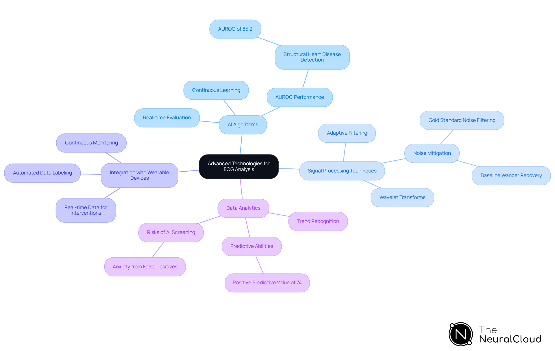
- AI Algorithms: MaxYield™ implements AI algorithms that enable real-time evaluation of ECG data, adeptly identifying patterns and anomalies that may escape human detection. Recent studies demonstrate that these AI models can achieve an area under the receiver operating characteristic curve (AUROC) of 85.2% in detecting structural heart disease (SHD), significantly surpassing traditional methods. This platform continuously evolves its algorithms with each use, over time.
- Signal Processing Techniques: The platform employs advanced signal processing methods, including wavelet transforms and adaptive filtering, to enhance the quality of ECG signals by effectively mitigating noise and artifacts. MaxYield™ excels in this domain, utilizing gold standard noise filtering and distinct wave recognition to recover previously obscured segments of recordings impacted by baseline wander and muscle artifact. These techniques have been proven to improve the clarity of ECG signals, leading to more accurate interpretations.
- Integration with Wearable Devices: MaxYield™ develops solutions that seamlessly integrate with wearable ECG monitors, enabling continuous monitoring and analysis of cardiac health. This integration not only boosts user engagement but also equips healthcare professionals with real-time data for timely interventions. The AI tool's ability to identify high-risk individuals for prompt echocardiography underscores the importance of real-time data in clinical decision-making. Additionally, MaxYield™ automates the labeling of critical data, streamlining workflows for developers.
- Data Analytics: Strong data analytics capabilities are utilized to compile and analyze extensive datasets from diverse individuals, facilitating trend recognition and enhancing predictive abilities. For instance, AI-enhanced ECG analysis has demonstrated a positive predictive value of 74% in identifying high-risk individuals, indicating the potential for improved patient outcomes. However, it is essential to address the risks associated with AI-based screening, such as anxiety from false positives, which requires further investigation. By leveraging the advanced analytics features of MaxYield™, including its data wall, developers can enhance platform functionality and reliability, ultimately contributing to better patient care and outcomes.
Conclusion
Understanding the intricate components of ECG waves is essential for developers aiming to enhance cardiac health monitoring tools. The P-wave, QRS complex, ST segment, and T-wave each play a pivotal role in depicting the heart's electrical activity, providing critical insights for diagnosing various cardiac conditions. A comprehensive grasp of these elements not only aids in accurate ECG interpretation but also fosters the development of innovative solutions that can significantly improve patient outcomes.
Key insights discussed include:
- The importance of standardization in ECG recordings
- The implementation of automated algorithms for efficient analysis
- The necessity of correlating ECG findings with clinical symptoms
Furthermore, the integration of advanced technologies such as AI algorithms and signal processing techniques exemplifies how developers can enhance the precision and reliability of ECG evaluations. These advancements empower healthcare professionals to make informed decisions swiftly, ultimately leading to better patient care.
In conclusion, the significance of mastering ECG wave components and interpretation techniques cannot be overstated. As developers continue to leverage cutting-edge technologies and methodologies, the potential for improved diagnostic accuracy and patient outcomes expands. By prioritizing education and innovation in ECG analysis, the healthcare community can ensure that cardiac health monitoring remains at the forefront of medical advancements, paving the way for more effective and timely interventions.
Frequently Asked Questions
What is an electrocardiogram (ECG)?
An electrocardiogram (ECG) is a graphical representation of the heart's electrical activity, used to assess cardiac function.
What does the P-wave signify in an ECG?
The P-wave signifies atrial depolarization, marking the electrical impulse that initiates atrial contraction, with a normal duration of approximately 80 milliseconds.
What is the significance of the QRS complex in an ECG?
The QRS complex represents ventricular depolarization and is crucial for effective heart pumping. Its duration is vital for evaluating ventricular function and should typically be less than 120 milliseconds.
What does the ST segment indicate in an ECG?
The ST segment indicates the interval between ventricular depolarization and repolarization, providing insights into the heart's recovery phase. Irregularities in this segment can indicate ischemia or other heart-related problems.
What is represented by the T-wave in an ECG?
The T-wave represents ventricular repolarization, illustrating the heart's return to its resting state. Its shape can signify various heart conditions.
What are the PR and QT intervals, and why are they important?
The PR interval (120-200 milliseconds) and QT interval (420 milliseconds or less at 60 bpm) are crucial for understanding conduction durations and identifying irregularities in heart conduction and function.
How does the MaxYield™ platform enhance ECG analysis?
The MaxYield™ platform enhances ECG analysis by utilizing advanced noise filtering and wave recognition techniques, enabling swift evaluation of ECG signals even amid artifacts and recovering hidden parts of recordings.
What are the benefits of using MaxYield™ for ECG evaluations?
MaxYield™ provides beat-by-beat evaluations of up to 200,000 heartbeats in under 5 minutes, improving diagnostic accuracy and supporting confident clinical decision-making.
What did Lawrence Rosenthal, MD, PhD, emphasize about tachycardias?
Lawrence Rosenthal emphasized that tachycardias generated from abnormal repolarization can result from genetic defects in ion channels (channelopathies) and can be lethal.
