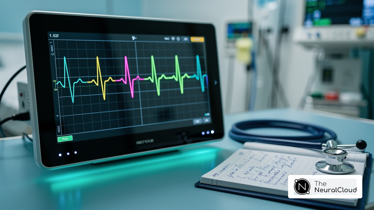
Introduction
Identifying conduction abnormalities like Right Bundle Branch Block (RBBB) and Left Anterior Fascicular Block (LAFB) in ECGs is a complex task for healthcare professionals. This guide provides a step-by-step approach to diagnosing these conditions, highlighting key characteristics and clinical implications that enhance patient care.
Healthcare professionals often struggle to accurately identify conduction abnormalities in ECGs due to the fast-paced nature of clinical environments. Misinterpretation can lead to incorrect diagnoses, impacting patient care and outcomes.
As the complexity of ECG interpretation increases, how can developers and clinicians work together to ensure accurate diagnoses?
Understand RBBB and LAFB: Key Characteristics and Implications
Identifying conduction abnormalities such as RBBB and LAFB through ECG analysis presents unique challenges for healthcare professionals.
Key Characteristics of RBBB:
- QRS Duration: Right Bundle Branch Block is characterized by a QRS duration surpassing 120 milliseconds, signifying a delay in ventricular conduction.
- 'RSR' Pattern: An RSR' pattern, often resembling an 'M' shape, is typically seen in positions V1-V3, indicating the presence of right bundle branch block.
- Wide S Wave: A wide, slurred S wave appears in lateral leads (I, aVL, V5, V6), further confirming the diagnosis.
- Clinical Implications: RBBB may suggest underlying heart conditions such as ischemic heart disease or structural abnormalities, necessitating further evaluation. MaxYield enhances the accuracy of detecting these conditions by reducing motion noise and providing consistent, clean data across devices, enabling beat-by-beat analysis and detailed insights.
Key Characteristics of LAFB:
- QRS Axis Deviation: Left anterior fascicular block is characterized by left axis deviation (LAD), which can be indicative of cardiac stress or pathology.
- QRS Morphology: The QRS complex typically presents a small R wave in lead I and a pronounced S wave in lead III, reflecting the altered conduction pattern.
- Clinical Implications: Left Anterior Fascicular Block serves as a potential marker for underlying cardiac issues, including hypertension and coronary artery disease, which may require clinical intervention. The advanced algorithms of MaxYield facilitate the identification of these abnormalities, enabling developers to create effective diagnostic tools that can accurately analyze ECG readings.
Understanding these characteristics can significantly enhance the development of diagnostic tools that accurately identify RBBB and LAFB, ultimately leading to improved patient care.
 and Left Anterior Fascicular Block (LAFB). Each branch represents a characteristic or clinical implication of these conditions. Follow the branches to explore how each characteristic contributes to understanding these conduction abnormalities.")
Perform ECG Analysis: Step-by-Step Diagnostic Process for RBBB and LAFB
To accurately diagnose Right Bundle Branch Block (RBBB) and Left Anterior Fascicular Block (LAFB) through ECG analysis, follow these steps:
Step 1: Obtain a 12-Lead ECG
- Ensure proper electrode placement to capture a comprehensive view of the heart's electrical activity. In Canada, adherence to established guidelines for electrode positioning is crucial for accurate readings.
Step 2: Analyze the QRS Complex
- Measure the QRS duration. A duration exceeding 120 ms indicates right bundle branch block, while a duration between 100 and 119 ms suggests incomplete right bundle branch block. Look for the characteristic RSR pattern in segments V1-V3, which is suggestive of right bundle branch block. Additionally, RBBB shows a slurred S wave in channels I, aVL, V5, and V6. Utilizing MaxYield™, you can enhance this analysis by leveraging its advanced noise filtering capabilities, which help isolate ECG waves even in recordings with significant baseline wander and muscle artifact.
Step 3: Assess the QRS Axis
- Determine the QRS axis using leads I and aVF. A left axis deviation indicates left anterior fascicular block, often associated with RBBB and LAFB, which is frequently linked to a small R wave in channel I and a deep S wave in channel III.
Step 4: Evaluate Additional Leads
- Check leads I, II, III, aVL, and aVF for characteristic changes associated with LAFB. The presence of a broad, dominant S wave in leads V1 and V2 is also a key indicator of RBBB and LAFB. MaxYield™ can assist in rapidly isolating these features, ensuring that critical data is not obscured by noise.
Step 5: Correlate with Clinical Findings
- Consider the patient's clinical history and symptoms. LBBB and LAFB may suggest underlying cardiac problems, such as hypertension or structural heart disease, that necessitate further examination. Keep in mind that right bundle branch block often shows no symptoms, making diagnosis trickier.
Step 6: Document Findings
- Record your findings clearly, noting any abnormalities and their potential clinical implications. This documentation is crucial for further analysis and treatment planning, ensuring that all relevant data is available for ongoing patient care. Remember that right bundle branch block can advance to issues like third-degree AV block, which may require more vigilant observation and action. Integrating MaxYield™ into your workflow enhances ECG analysis clarity and efficiency, improving patient outcomes.

Integrate Advanced Tools: Enhance ECG Diagnostics with AI Technology
To enhance ECG diagnostics for RBBB and LAFB, consider integrating the following advanced tools:
- MaxYield: Leverage Neural Cloud Solutions Inc.'s MaxYield platform, which utilizes patented signal mapping algorithms to effectively clean and isolate ECG signals. This significantly improves diagnostic accuracy, especially in detecting subtle waveform changes related to rbbb and lafb. MaxYield is great at reducing noise, quickly isolating ECG waves from recordings that might have baseline wander, movement, or muscle artifacts. This means that even lengthy Holter, 1-Lead, and patch monitor recordings can still provide critical data. Additionally, MaxYield delivers beat-by-beat analysis and identifies cardiac events, enhancing the overall diagnostic process.
- Insight360: Employ this visualization tool to create customizable dashboards and reports, facilitating better trend analysis and data interpretation. By visualizing ECG data effectively, healthcare professionals can make more informed decisions regarding patient care.
Implementation Steps:
- Select Compatible Devices: Ensure that the ECG devices in use are compatible with AI tools to maximize integration efficiency.
- Integrate with Existing Workflows: Seamlessly incorporate AI tools into current ECG analysis workflows to minimize disruption and enhance operational efficiency.
- Train Staff: Provide comprehensive training for medical professionals on effectively utilizing AI tools, focusing on interpreting AI-generated insights to improve diagnostic accuracy.
- Monitor Performance: Regularly evaluate the efficacy of AI tools in diagnosing rbbb and lafb, making necessary adjustments to enhance accuracy and reliability.
Detecting subtle waveform changes in ECG analysis can be challenging, often leading to misdiagnoses. By leveraging AI technology, developers can significantly enhance the diagnostic capabilities of ECG analysis, leading to improved patient outcomes. The integration of AI-powered tools has shown to increase diagnostic sensitivity and specificity, with studies indicating that AI-ECG can achieve a sensitivity of 92% and specificity of 81% compared to traditional methods. Notably, AI-ECG demonstrated a 99.1% negative predictive value, confirming that most patients without LVSD were accurately identified. Furthermore, AI-ECG has shown a remarkable 91% reduction in false-positive activations, decreasing from 41.8% to 7.9% for patients with negative cardiac biomarkers. The integration of AI-powered tools not only streamlines the diagnostic process but also empowers healthcare professionals to provide timely and accurate care, ultimately improving patient outcomes.

Troubleshoot Common Issues: Overcoming Challenges in ECG Diagnosis
When diagnosing RBBB and LAFB, developers often encounter challenges that can complicate accurate ECG analysis. Here’s how to troubleshoot them:
Issue 1: Inconsistent QRS Morphology
- Solution: Make sure the electrodes are placed correctly and look out for any movement artifacts. Re-run the ECG if necessary. Signal artifacts and electrical interference can distort ECG readings, so maintaining a static-free environment is crucial for enhancing signal quality.
Issue 2: Misinterpretation of Axis Deviation
- Solution: Take a moment to double-check the QRS axis calculation. Use multiple leads to confirm findings and consider the patient's clinical context. Studies indicate that incorrect diagnoses of ischemia can correlate with factors such as body mass index (BMI), which may influence interpretation accuracy.
Issue 3: Overlapping Conditions
- Solution: Keep in mind that other conditions might look similar to RBBB or LAFB. Use additional diagnostic tools, such as echocardiography, to clarify ambiguous cases. Automated ECG diagnostic devices have shown a wide range of misinterpretations, with incorrect diagnoses for ST-elevation myocardial infarction (STEMI) ranging from 0% to 42%, highlighting the importance of thorough evaluation.
Issue 4: AI Tool Limitations
- Solution: It's important to recognize the limitations of AI tools. Regularly update the algorithms and ensure that staff are trained to interpret AI outputs critically. A study found that 39% of ECGs were interpreted incorrectly, emphasizing the need for clinician verification to avoid misinterpretations that could mislead medical staff.
By proactively addressing these challenges, developers can significantly enhance diagnostic accuracy and ultimately improve patient care outcomes.

Conclusion
Diagnosing Right Bundle Branch Block (RBBB) and Left Anterior Fascicular Block (LAFB) presents unique challenges for healthcare professionals. Understanding the characteristics of RBBB and LAFB and using a systematic ECG interpretation approach can significantly enhance diagnostic accuracy and patient outcomes.
This article outlines a step-by-step diagnostic process that begins with obtaining a 12-lead ECG and analyzing the QRS complex for specific patterns indicative of RBBB and LAFB. It highlights the importance of integrating advanced tools like MaxYield and Insight360, which improve ECG diagnostics by reducing noise and enhancing data clarity. Additionally, it addresses common challenges encountered during ECG interpretation and offers practical solutions to overcome these obstacles.
With advanced AI tools in ECG diagnostics, healthcare professionals can analyze data more efficiently and make quicker, informed decisions. By prioritizing accurate diagnoses of RBBB and LAFB, the medical community can better address underlying cardiac issues, leading to improved patient care and outcomes. Adopting these advanced methodologies will not only enhance diagnostic accuracy but also transform patient care in cardiology.
Frequently Asked Questions
What is Right Bundle Branch Block (RBBB)?
Right Bundle Branch Block (RBBB) is a conduction abnormality characterized by a QRS duration exceeding 120 milliseconds, indicating a delay in ventricular conduction.
How can RBBB be identified on an ECG?
RBBB can be identified by an 'RSR' pattern resembling an 'M' shape in leads V1-V3 and a wide, slurred S wave in lateral leads (I, aVL, V5, V6).
What are the clinical implications of RBBB?
RBBB may indicate underlying heart conditions such as ischemic heart disease or structural abnormalities, which require further evaluation.
What is Left Anterior Fascicular Block (LAFB)?
Left Anterior Fascicular Block (LAFB) is characterized by left axis deviation (LAD), which can suggest cardiac stress or pathology.
How can LAFB be identified on an ECG?
LAFB is identified by a small R wave in lead I and a pronounced S wave in lead III, reflecting an altered conduction pattern.
What are the clinical implications of LAFB?
LAFB may serve as a marker for underlying cardiac issues, including hypertension and coronary artery disease, which may necessitate clinical intervention.
How does MaxYield enhance the detection of RBBB and LAFB?
MaxYield improves the accuracy of detecting RBBB and LAFB by reducing motion noise and providing consistent, clean data across devices, enabling detailed beat-by-beat analysis.
List of Sources
- Understand RBBB and LAFB: Key Characteristics and Implications
- litfl.com (https://litfl.com/bifascicular-block-ecg-library)
- thecardiologyadvisor.com (https://thecardiologyadvisor.com/ddi/right-bundle-branch-block-rbbb)
- my.clevelandclinic.org (https://my.clevelandclinic.org/health/diseases/23212-left-anterior-fascicular-block)
- ecgweekly.com (https://ecgweekly.com/ecgstat/new-rbbb-lafb)
- Perform ECG Analysis: Step-by-Step Diagnostic Process for RBBB and LAFB
- thecardiologyadvisor.com (https://thecardiologyadvisor.com/ddi/right-bundle-branch-block-rbbb)
- rebelem.com (https://rebelem.com/bundle-branch-blocks101)
- Checking your browser - reCAPTCHA (https://pmc.ncbi.nlm.nih.gov/articles/PMC8142372)
- litfl.com (https://litfl.com/bifascicular-block-ecg-library)
- Integrate Advanced Tools: Enhance ECG Diagnostics with AI Technology
- AI-ECG Finds STEMI Faster, Cuts False-Positive Cath Lab Activations (https://tctmd.com/news/ai-ecg-finds-stemi-faster-cuts-false-positive-cath-lab-activations)
- Study Shows AI-Powered ECG Could Help Patients Requiring Lifelong Heart Monitoring (https://dicardiology.com/content/study-shows-ai-powered-ecg-could-help-patients-requiring-lifelong-heart-monitoring)
- AI-ECG Momentum and Rising Competition Reshape Diagnostic Cardiology in 2026 (https://signifyresearch.net/insights/diagnostic-cardiology-market-bouncing-into-2026)
- AI-based ECG interpretation outperforms standard diagnosis of occlusive myocardial infarction (https://news-medical.net/news/20260323/AI-based-ECG-interpretation-outperforms-standard-diagnosis-of-occlusive-myocardial-infarction.aspx)
- AI-powered electrocardiogram detects early signs of heart failure (https://utsouthwestern.edu/newsroom/articles/year-2026/may-ai-powered-electrocardiogram.html)
- Troubleshoot Common Issues: Overcoming Challenges in ECG Diagnosis
- allstatesmed.com (https://allstatesmed.com/blogs/news/how-to-troubleshoot-common-ecg-machine-failures?srsltid=AfmBOoo-xaMnYsIVsN3-dcz_WhCQ2CfJjN-spdxStCrNoiDfSZAgZA-V)
- Checking your browser - reCAPTCHA (https://pmc.ncbi.nlm.nih.gov/articles/PMC12137353)
- Frontiers | The most common errors in automatic ECG interpretation (https://frontiersin.org/journals/physiology/articles/10.3389/fphys.2025.1590170/full)